

Case Reports
doi: 10.3941/jrcr.v6i7.1076.
Epub 2012 Jul 1.
Ureteral fibroepithelial polyp causing urinary obstruction
Affiliations
- PMID: 23365709
- PMCID: PMC3558047
- DOI: 10.3941/jrcr.v6i7.1076
Item in Clipboard
Case Reports
Ureteral fibroepithelial polyp causing urinary obstruction
J Radiol Case Rep.
2012 Jul.
Abstract
Ureteral polyps are rare causes of ureteropelvic junction (UPJ) obstruction, particularly in children. We report a nine year-old boy with UPJ obstruction initially suggestive of an obstructive urinary stone. CT showed intraureteral calcification at the UPJ and hydronephrosis. A retrograde pyelogram showed narrowing at the UPJ and partial obstruction that was found to be a ureteral polyp. This case illustrates a rare cause of UPJ obstruction that should be considered when the imaging findings and presentation are atypical for more common etiologies of ureteral obstruction.
Keywords: fibroepithelial polyp; pediatric; ureteral polyp; ureteropelvic junction obstruction.
Figures
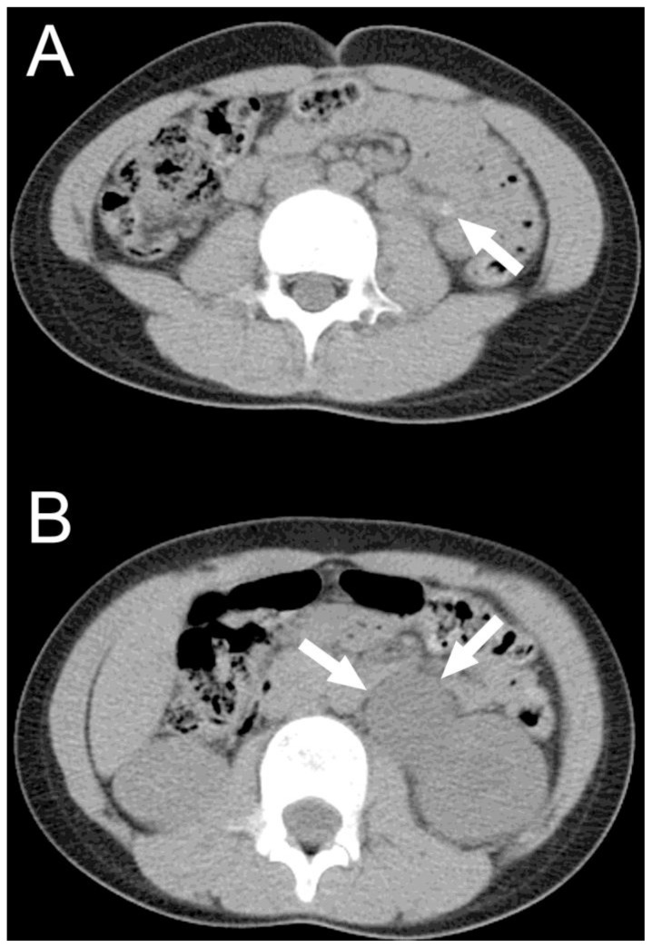
9 year-old boy with ureteropelvic junction obstruction found to be caused by a 2cm ureteral polyp. A transverse, non-contrast CT scan shows a calcified ureteral polyp (arrow in A) in the patient’s left ureter, causing proximal obstruction of the renal pelvis (arrows in B). Non-contrast CT of the abdomen and pelvis was obtained on a GE LightSpeed VCT at mA=109–139 with automatic current modulation, kvp=120 and reconstructed into 5 mm thick axial images.

9 year-old boy with a ureteropelvic junction obstruction found to be caused by a 2cm ureteral polyp. A coronal, non-contrast CT scan shows a ureteral polyp with calcification (arrow) in the patient’s left ureter, causing proximal obstruction of the renal pelvis and hydronephrosis. Non-contrast CT of the abdomen and pelvis was obtained on a GE LightSpeed VCT at mA=109–139 with automatic current modulation, kvp=120 and reconstructed into 5 mm thick coronal images.

9 year-old boy with ureteropelvic junction obstruction found to be caused by a 2cm ureteral polyp. A) Grayscale renal ultrasound shows grade III hydronephrosis. B) Renal Doppler ultrasound shows blood flow in the kidney, proximal urinary obstruction, and the obstructing hypervascular mass (arrow). Doppler image obtained with a curved transducer at 5 MHz on an Acuson imaging system.

9 year-old boy with a ureteropelvic junction obstruction found to be caused by a 2cm ureteral polyp. MAG3 lasix renogram shows decreased function in the left kidney is compatible with urinary obstruction.

9 year-old boy with ureteropelvic junction obstruction found to be caused by a 2 cm ureteral polyp. A retrograde pyelogram of the left proximal ureter showed irregularity and stenosis (arrows) but did not reveal a clear etiology for the obstruction. Spot fluoroscopic image was obtained at 69 kVp and 2.12 mA after the retrograde injection of non-ionic water-soluble contrast material.
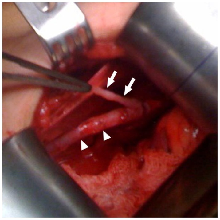
9 year-old boy with ureteropelvic junction obstruction found to be caused by a ureteral polyp. Intraoperative photo shows the ureter (arrow heads) and 2cm long stalk of the polyp (arrows)
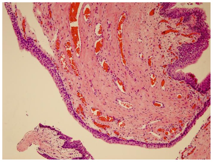
9 year-old boy with ureteropelvic junction obstruction found to be a ureteral polp. The image is at 200× magnification with a hematoxylin and eosin stain. Pathology showed a fibroepithelial polyp demonstrating a thin, bland urothelial cell lining over a polypoid fibrovascular stroma with frequent dilated and congested small blood vessels
References
-
- Adey GS, Vargas SO, Retik AB, Borer JG, Mandell J, Hendren WH, Lebowitz RL, Bauer SB. Fibroepithelial polyps causing ureteropelvic junction obstruction in children. J Urol. 2003 May;169(5):1834–6. - PubMed
-
- Niu ZB, Yang Y, Hou Y, Chen H, Wang CL. Ureteral polyps: an etiological factor of hydronephrosis in children that should not be ignored. Pediatr Surg Int. 2007 Apr;23(4):323–6. - PubMed
-
- Lavelle JP, Knisely AS, Bellinger MF. Benign fibroepithelial polyps causing symptomatic bilateral intermittent hydroureteronephrosis. J Urol. 1997 Aug;158(2):569. - PubMed
-
- Bartone FF, Johansson SL, Markin RJ, Imray TJ. Bilateral fibroepithelial polyps of ureter in a child. Urology. 1990 Jun;35(6):519–22. - PubMed
-
- Romesburg JW, Stein RJ, Desai MM, Lagwinski N, Ross JH. Treatment of child with bilateral ureteropelvic junction obstruction due to fibroepithelial polyps and review of the literature. Urology. 2009 Apr;73(4):929 e9–11. - PubMed