

Case Reports
doi: 10.3941/jrcr.v6i8.1027.
Epub 2012 Aug 1.
Paraperitoneal inguinal hernia of ureter
Affiliations
- PMID: 23365714
- PMCID: PMC3558268
- DOI: 10.3941/jrcr.v6i8.1027
Item in Clipboard
Case Reports
Paraperitoneal inguinal hernia of ureter
J Radiol Case Rep.
2012 Aug.
Abstract
Inguinal herniation of ureter is an uncommon finding that can potentially lead to obstructive uropathy. We report a case of inguinal herniation of ureter discovered incidentally during workup for acute renal failure and ultrasound finding of hydronephrosis.
Keywords: Computed tomography; genitourinary.
Figures
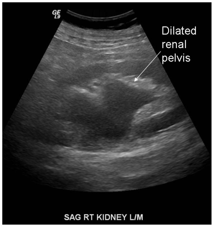
Dilated renal pelvis. Sagittal sonographic image was obtained with a 4 curved transducer at 4.0 MHz of the right kidney from a 76 year old male with acute renal failure, demonstrating dilated right renal pelvis and proximal ureter.

Extra renal pelvis and inguinal herniation of the right ureter. The patient is a 76 year old male presenting with acute renal failure. Here is an oblique reconstructed CT image showing a dilated renal pelvis with intact renal pelvic fat and gradual tapering of the proximal ureter consistent with an extrarenal pelvis. The ureter is seen going into the inguinal canal. There is no evidence of ureteral obstruction. This study was performed using GE LightSpeed 16-slice CT scanner using kV 140 and mA 379. 40 cc in intravenous Visipaque was injected at time 0. At 5 minutes an additional 120 cc of Visipaque was injected. At 7 minutes a combined nephrographic/execretory phase helical CT was performed of the abdomen and pelvis using 0.625 mm collimation with 2.5 mm axial reconstructions and 3 mm bilateral oblique reconstructions.

Inguinal hernia of the ureter. The patient is a 76 year old male presenting with acute renal failure. Axial image of CT urogram with the patient in a prone position shows a contrast-filled right ureter within the right inguinal canal. Bilateral inguinal hernia sacs are present, and the herniated ureter is seen medial to the right inguinal hernia sac. Bilateral hip prostheses are present. This study was performed using GE LightSpeed 16-slice CT scanner using kV 140 and mA 379. 40 cc in intravenous Visipaque was injected at time 0. At 5 minutes an additional 120 cc of Visipaque was injected. At 7 minutes a combined nephrographic/execretory phase helical CT was performed of the abdomen and pelvis using 0.625 mm collimation with 2.5 mm axial reconstructions and 3 mm bilateral oblique reconstructions.

Inguinal hernia of the ureter. The patient is a 76 year old male presenting with acute renal failure. 3-dimensional reconstruction image of CT urogram showing the right ureter going beneath the pelvic rim before making a hairpin turn up towards the bladder. This study was performed using GE LightSpeed 16-slice CT scanner using kV 140 and mA 379. 40 cc in intravenous Visipaque was injected at time 0. At 5 minutes an additional 120 cc of Visipaque was injected. At 7 minutes a combined nephrographic/execretory phase helical CT was performed of the abdomen and pelvis using 0.625 mm collimation with 2.5 mm axial reconstructions and 3 mm bilateral oblique reconstructions.

Inguinal hernia of the ureter. The patient is a 76 year old male presenting with acute renal failure. 3-dimensional reconstruction image of CT urogram showing the right ureter going beneath the pelvic rim before making a hairpin turn up towards the bladder. This study was performed using GE LightSpeed 16-slice CT scanner using kV 140 and mA 379. 40 cc in intravenous Visipaque was injected at time 0. At 5 minutes an additional 120 cc of Visipaque was injected. At 7 minutes a combined nephrographic/execretory phase helical CT was performed of the abdomen and pelvis using 0.625 mm collimation with 2.5 mm axial reconstructions and 3 mm bilateral oblique reconstructions.

The patient is a 76 year old male presenting with acute renal failure. Axial CT image shows placement of a percutaneous drain into a left renal abscess. Contrast excretion into the right renal pelvis was seen from previously administered contrast from a different study. This study was performed using GE LightSpeed 16-slice CT scanner using kV 140 and mA 379. No intravenous contrast was administered for this study.
References
-
- Odisho A, et al. Inguinal herniation of a transplant ureter. Kidney International. 2010;78:115. - PubMed
-
- Zarraonandia Andraca A, et al. Inguinal ureteral hernia: a clinical case. Arch Esp Urol. 2009;62(9):755–757. - PubMed
-
- Schlussel RN, Retik AB. Ectopic ureter, ureterocele, and other anomalies of the ureter. In: Walsh PC, Wein AJ, Vaughan ED, et al., editors. Campbell’s urology. 8th ed. Oxford: WB Saunders; 2002. pp. 2047–8.
-
- Roach SC, Moulding F, Hanbidge A. Inguinal herniation of the ureter. Am J Roentgenol. 2005 Jul;185(1):283. - PubMed
-
- Bertolaccini L, Giacomelli G, Bozzo RE, Gastaldi L, Moroni M. Inguinaoscrotal hernia of a double district ureter: case report and literature review. Hernia. 2005;9:291–293. - PubMed