Case Reports
. 2012 Aug;6(8):27-38.
doi: 10.3941/jrcr.v6i8.1052. Epub 2012 Aug 1.

Presentation of ileal Burkitt lymphoma in children

Affiliations
Case Reports

Presentation of ileal Burkitt lymphoma in children

Joseph R Grajo et al. J Radiol Case Rep. 2012 Aug.

Abstract

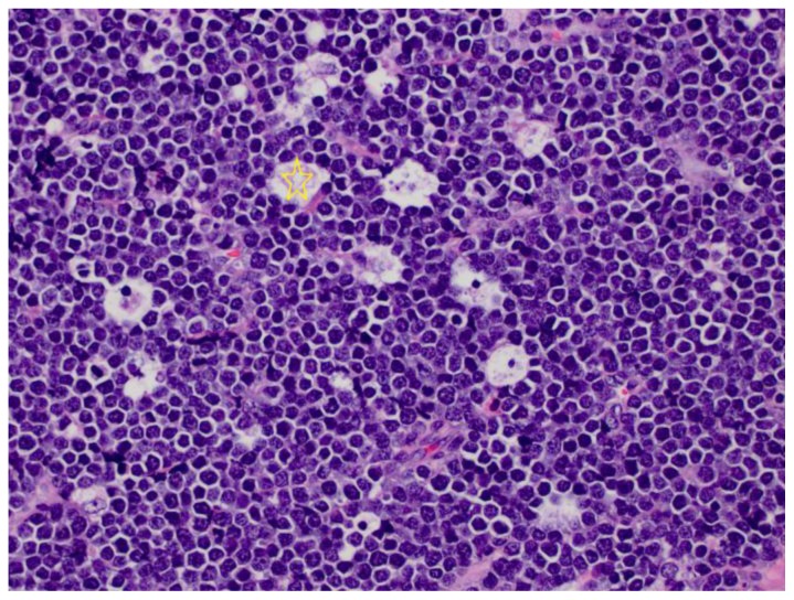
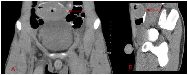
Burkitt lymphoma is a highly aggressive form of Non-Hodgkin lymphoma that responds favorably if diagnosed accurately and treated early. Recognition of the various radiologic manifestations of Burkitt lymphoma can help guide the clinician to expedite appropriate chemotherapy. We present two cases that illustrate different radiologic presentations of this aggressive gastrointestinal malignancy in children. Case 1 features a 7-year-old boy who presented to our hospital with recurrent ileocecal intussusception. Case 2 describes a 16-year-old male with history of blood-streaked stools. Ileocectomy was performed in both cases and histologic analysis showed the "starry sky pattern" and t(8;14) translocation, classic for Burkitt lymphoma. Both patients remain disease-free following surgical excision and chemotherapy.

Keywords: Burkitt; gastrointestinal; lymphoma; pediatric.


Figures

Figure 1
Figure 1
7-year-old Caucasian male with ileal Burkitt lymphoma. Focused sonography of the right lower quadrant performed at initial presentation demonstrates a reniform-appearing mass in the right lower quadrant, suspicious for intussusception (arrow, A) with free fluid in the right lower quadrant (arrow, B). Minimal color flow is seen (C). (Protocol: GE M12L transducer with pediatric abdomen settings at 9 MHz frequency. Color flow images obtained at 5 MHz)
Figure 2
Figure 2
7-year-old Caucasian male with ileal Burkitt lymphoma. Axial (A) and coronal (B) CT images of the abdomen and pelvis demonstrate a typical target-appearing lesion in the right lower quadrant, suggestive of intussusception of the terminal ileum into the cecum (arrows) with free fluid in the right paracolic gutter. (Protocol: Phillips Brilliance 64 slice CT scanner, 78 mA, 80 kV, 3 mm slice thickness, CTDIvol 0.842 mGy, 50 mL Optiray 350 IV contrast at 1 mL/s, 360 mL rectal contrast, no oral contrast)
Figure 3
Figure 3
7-year-old Caucasian male with ileal Burkitt lymphoma. Following initial diagnostic work-up, the patient was taken for air contrast enema. Scout (A), supine (B), and upright (C) views are provided. Residual oral contrast from the CT was seen in the transverse and descending colon. The ileocecal intussusception was successfully reduced by the radiologist. A lobulated wall-based soft tissue density at the ileocecal junction (arrows) was initially felt to represent an edematous ileocecal valve. In retrospect, this most likely represented the mass. (Protocol: Soft tip catheter placed intra-rectally and taped into position. Using fluoroscopic guidance, a sphygmometer was used with 120 mmHg pop off for maximum air pressure. Air was pumped into the colon for 5 minutes.)
Figure 4
Figure 4
7-year-old Caucasian male with ileal Burkitt lymphoma. Focused sonography of the right lower quadrant demonstrates a “target sign,” consistent with ileocecal intussusception (arrow). At this point, the patient was taken to the operating room for laparotomy with eventual ileocecectomy. (Protocol: GE 9L transducer with pediatric abdomen settings at 8 MHz frequency)
Figure 5
Figure 5
7 year-old Caucasian male with ileal Burkitt lymphoma. Fleshy exophytic tumor (stars) involves most of the circumference of the ileocecal valve and bulges into the cecal lumen.
Figure 6
Figure 6
7-year-old Caucasian male with ileal Burkitt lymphoma. The tumor is composed of monomorphic, intermediate size lymphocytes with “mosaic tile” molding of the cells, and “starry sky” pattern, with large tingible body macrophages containing cellular debris (star).
Figure 7
Figure 7
16-year-old Hispanic male with ileal Burkitt lymphoma. Upright abdominal radiograph at initial presentation demonstrates a nonspecific bowel gas pattern with gaseous distention of several small bowel loops (arrow).
Figure 8
Figure 8
16-year-old Hispanic male with ileal Burkitt lymphoma. Axial (A), coronal (B), and sagittal (C), contrasted CT of the abdomen/pelvis demonstrate an 8 cm mass at the level of the pelvic inlet with central hypoattenuation and gas, concerning for necrosis and abscess/ulceration (arrows). Further evaluation with delayed imaging was recommended. (Protocol: Phillips Brilliance 64 slice CT scanner, 78 mA, 120 kV, 3 mm slice thickness, CTDIvol 6.692 mGy, 110 mL Optiray 350 IV contrast at 2.5 mL/s, oral contrast)
Figure 9
Figure 9
16-year-old Hispanic male with ileal Burkitt lymphoma. Axial (A) and sagittal (B) delayed CT images were obtained through the pelvis six hours following the initial CT. This exam demonstrates the large pelvic mass outlined by oral contrast (arrows), suggesting intraluminal extension and invasion into the small bowel. The area of central necrosis is better delineated and correlates with pathology as described in Figure 11. (Protocol: Phillips Brilliance 64 slice scanner, 99 mA, 120 kV, 3 mm slice thickness, CTDIvol 4.875 mGy, 6 hour delay following original CT)
Figure 10
Figure 10
16-year-old Hispanic male with ileal Burkitt lymphoma. A large ileal tumor with overlying hemorrhagic serosa and focal disruption (star).
Figure 11
Figure 11
16-year-old Hispanic male with ileal Burkitt lymphoma. Bisected specimen shows a fleshy tumor bulging into the ileal lumen, with extensive hemorrhage and central necrosis, outlined by the red arrows. Irregularity in the serosal surface is also demonstrated (on the left). The ileocecal valve (ICV) was not involved.
Figure 12
Figure 12
16-year-old Hispanic male with ileal Burkitt lymphoma. Three days following surgery and pathologic diagnosis, the patient received a whole body bone scan to evaluate for osseous metastasis. Anterior and posterior views of the raw and truncated data are shown. No bony metastasis was identified. (Protocol: Picker Prism 2000 Dual Head Nuclear Gamma Camera with medium energy collimator, Radiotracer − 17.1 mCi Tc99m MDP intravenously, imaged 3 hours after administration)
Figure 13
Figure 13
16-year-old Hispanic male with ileal Burkitt lymphoma. Following whole body bone scan, the patient was imaged for a gallium scan to evaluate for residual tumor or extra deposits of Burkitt lymphoma not already identified. Images were obtained at 72, 120, and 168 hours following administration of Ga67 citrate. A spot LAO view at 72 hours and LAO SPECT image at 120 hours show intense activity in the left upper pelvis. It was thought that this may represent postsurgical, inflammatory changes but possibility of residual disease was considered given intense focal radiotracer uptake. (Protocol: Picker Prism 2000 Dual Head Nuclear Gamma Camera with medium energy collimator, 7 mCi Ga67 citrate via left chest Mediport; images obtained at 72, 120, and 168 hours following injection; Whole body, spot, and SPECT images were obtained)
Figure 14
Figure 14
16-year-old Hispanic male with ileal Burkitt lymphoma. Follow up CT and Ga67 scans were obtained after chemotherapy at a 9 month interval following surgical resection. Both studies demonstrated complete resolution of disease. Selected axial (A) and coronal (B) CT images of the abdomen/pelvis show complete resection of the mass without evidence of residual or recurrent disease. (Protocol: Phillips Brilliance 64 slice scanner, 114 mA, 120 kV, 3 mm slice thickness, CTDIvol 6.212 mGy, 100 mL Optiray 350 IV contrast, oral contrast). Selected anterior and posterior whole body gallium scan at 120 hours (C) and selected coronal SPECT images at 168 hours (D) show complete resolution of postsurgical increased radiotracer uptake. (Protocol: Picker Prism 2000 Dual Head Nuclear Gamma Camera with medium energy collimator, 7.7 mCi Ga67 citrate intravenously, imaged at 120 and 168 hours following radiotracer injection)

References

    1. Gupta H, Davidoff AM, Pui CH, Shochat SJ, Sandlund JT. Clinical implications and surgical management of intussusception in pediatric patients with Burkitt lymphoma. J Pediatr Surg. 2007;42(6):998–1001. - PubMed
    1. Sorantin E, Lindbichler F. Management of intussusception. Eur Radiol. 2004;14(Suppl 4):L146–154. - PubMed
    1. Linabery AM, Ross JA. Childhood and adolescent cancer survival in the US by race and ethnicity for the diagnostic period 1975–1999. Cancer. 2008;113:2575–2596. - PMC - PubMed
    1. Kassira N, Pedroso FE, Cheung MC, Koniaris LG, Sola JE. Primary gastrointestinal tract lymphoma in the pediatric patient: review of 265 patients from the SEER registry. J Pediatr Surg. 2011;46:1956–1964. - PubMed
    1. Ferry JA. Burkitt’s lymphoma: clinicopathologic features and differential diagnosis. Oncologist. 2006;11(4):375–383. - PubMed

Publication types

MeSH terms