

Case of the diffuse form of xanthogranulomatous pyelonephritis
- PMID: 23365780
- PMCID: PMC3556416
- DOI: 10.1155/2013/936035
Case of the diffuse form of xanthogranulomatous pyelonephritis
Abstract
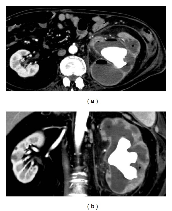
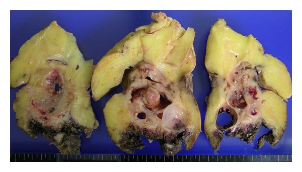
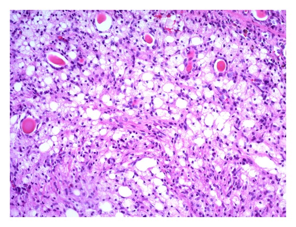
Xanthogranulomatous pyelonephritis (XGP) is a rare entity and constitutes less than 1% of chronic pyelonephritis. A 71-year-old male was introduced to our department with general malaise and abnormal findings of computed tomography (CT). Abnormal findings of complete blood count and laboratory examination included an elevated WBC count and C-reactive protein. Urinalysis showed combined hematuria and pyuria, and Escherichia coli was detected in urine culture. Abdominal CT revealed left hydronephrosis with staghorn renal calculi and thin cortex of the left kidney. Because of poor condition, the patient underwent construction of the left nephrostomy. After that, the remission of the inflammation was achieved. Several months after the construction, frequent obstructions of nephrostomy catheter because of turbid urine and intermittent fever elevation were observed. The patient and his family desired left nephrectomy despite his poor condition in general. Surgical dissection was very difficult due to fixed mass. Not long after that the patient died due to sepsis and cardiovascular failure. Microscopic findings of the left kidney revealed infiltration of lymphocytes and lipid-laden macrophages (xanthoma cells) corresponding to XGP.
Figures
Similar articles
-
Xanthogranulomatous pyelonephritis in children: diagnostic and therapeutic aspects.Eur J Pediatr Surg. 2002 Feb;12(1):42-8. doi: 10.1055/s-2002-25085. Eur J Pediatr Surg. 2002. PMID: 11967759
-
Xanthogranulomatous pyelonephritis with calculus migration into the psoas abscess: an unusual complication.Autops Case Rep. 2020 Dec 8;11:e2020200. doi: 10.4322/acr.2020.200. eCollection 2021. Autops Case Rep. 2020. PMID: 34277481 Free PMC article.
-
Xanthogranulomatous pyelonephritis: clinical experience with 41 cases.Urology. 2008 Feb;71(2):178-80. doi: 10.1016/j.urology.2007.09.026. Urology. 2008. PMID: 18308077 Review.
-
[Xanthogranulonatous pyelonephritis: report of 5 cases].Beijing Da Xue Xue Bao Yi Xue Ban. 2018 Aug 18;50(4):743-746. Beijing Da Xue Xue Bao Yi Xue Ban. 2018. PMID: 30122783 Chinese.
-
[A clinical study of xanthogranulomatous pyelonephritis (XGP) with special emphasis on the magnetic resonance imaging].Hinyokika Kiyo. 1992 Jan;38(1):43-6. Hinyokika Kiyo. 1992. PMID: 1546568 Review. Japanese.
Cited by
-
A Very Rare Case of Primary Renal Squamous Cell Carcinoma Presenting With Xanthogranulomatous Pyelonephritis on Radiological Imaging.Cureus. 2020 Dec 3;12(12):e11871. doi: 10.7759/cureus.11871. Cureus. 2020. PMID: 33282610 Free PMC article.
-
Clinicopathological Spectrum of Xanthogranulomatous Pyelonephritis.Indian J Nephrol. 2019 Mar-Apr;29(2):111-115. doi: 10.4103/ijn.IJN_50_18. Indian J Nephrol. 2019. PMID: 30983751 Free PMC article.
-
Nephron-sparing management of Xanthogranulomatous pyelonephritis presenting as spontaneous renal hemorrhage: a case report and literature review.BMC Urol. 2018 Jun 5;18(1):57. doi: 10.1186/s12894-018-0354-3. BMC Urol. 2018. PMID: 29866085 Free PMC article. Review.
-
Xanthogranulomatous Pyelonephritis with Staghorn Calculus, Acute Gangrenous Appendicitis and Enterocolitis: A Multidisciplinary Challenge of Kidney-Preserving Conservative Therapy.Curr Urol. 2015 Sep;8(3):162-5. doi: 10.1159/000365709. Epub 2015 Sep 4. Curr Urol. 2015. PMID: 26889137 Free PMC article.
-
Fluorodeoxyglucose positron emission tomography-computed tomography findings in a case of xanthogranulomatous pyelonephritis.Indian J Nucl Med. 2013 Jan;28(1):49-50. doi: 10.4103/0972-3919.116809. Indian J Nucl Med. 2013. PMID: 24019680 Free PMC article.
References
-
- Petronic V, Buturovic J, Isvaneski M. Xanthogranulomatous pyelonephritis. British Journal of Urology. 1989;64(4):336–338. - PubMed
-
- Eastham J, Ahlering T, Skinner E. Xanthogranulomatous pyelonephritis: clinical findings and surgical considerations. Urology. 1994;43(3):295–299. - PubMed
-
- Schlangenhaufer F. Uber eingentumiche Staphylomykosen der nieren und des pararennalen Bindewebes. Frankfurter Zeitschrift für Pathologie. 1916;19:139–148.
-
- Bazeed MA, Nabeeh A, Atwan N. Xanthogranulomatous pyelonephritis in bilharzial patients: a report of 25 cases. The Journal of Urology. 1989;141(2):261–264. - PubMed
-
- Mhiri MN, Sellami F, Njeh M, Rebai T, Smida ML. Xanthogranulomatous pyelonephritis: apropos of 42 cases. Annales d’Urologie. 1991;25(3):113–117. - PubMed
LinkOut - more resources
Full Text Sources
Other Literature Sources
Research Materials
