. 2012:2012:1358-61.
doi: 10.1109/EMBC.2012.6346190.

Implementation of topographically constrained connectivity for a large-scale biologically realistic model of the hippocampus

Affiliations

Implementation of topographically constrained connectivity for a large-scale biologically realistic model of the hippocampus

Gene J Yu et al. Annu Int Conf IEEE Eng Med Biol Soc. 2012.

Abstract

In order to understand how memory works in the brain, the hippocampus is highly studied because of its role in the encoding of long-term memories. We have identified four characteristics that would contribute to the encoding process: the morphology of the neurons, their biophysics, synaptic plasticity, and the topography connecting the input to and the neurons within the hippocampus. To investigate how long-term memory is encoded, we are constructing a large-scale biologically realistic model of the rat hippocampus. This work focuses on how topography contributes to the output of the hippocampus. Generally, the brain is structured with topography such that the synaptic connections formed by an input neuron population are organized spatially across the receiving population. The first step in our model was to construct how entorhinal cortex inputs connect to the dentate gyrus of the hippocampus. We have derived realistic constraints from topographical data to connect the two cell populations. The details on how these constraints were applied are presented. We demonstrate that the spatial connectivity has a major impact on the output of the simulation, and the results emphasize the importance of carefully defining spatial connectivity in neural network models of the brain in order to generate relevant spatiotemporal patterns.


Figures

Figure 1
Figure 1
Summary of the bands presented by Dolorfo and Amaral. Cells from the bands in the EC map to the corresponding colored areas on the DG. The 2-D postsynaptic absolute map of the DG is depicted.
Figure 2
Figure 2
Symbolic representation of the spatial connectivity mapping EC cells (left) to the DG (right) with a uniform random distribution. The absolute map of the EC is reduced to a 1-D array and is represented by the red bar with the first cell at the top and the last cell at the bottom. The lengths of the blue boxes symbolize the proportion of cells that map to a certain quartile. Overlap occurs in the darker blue boxes, and those cells can map to either quartile that contributes to the band (bottom). The sizes of the boxes do not represent the actual percentages used by the model and are for demonstrative purposes.
Figure 3
Figure 3
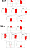
Validation of the connectivity matrix implementation. The subsets of cells that belonged to a particular band or overlap region in the EC were activated. The red dots signify the location of the granule cells that fired an action potential over 50 ms. Activity beyond the quartiles can be seen only the perforation points, not the axon terminal fields were constrained by the quartiles.
Figure 4
Figure 4
Output of the networks using the topographically constrained connectivity (left) and using random connectivity (right). Each point represents an action potential being fired at that time. The MEA and LEA cells are plotted by their cell ID, and the granule cells are plotted versus based on their septotemporal position on the DG. Only 500 ms of the simulation are shown in order to keep the plots from being too dense.

Similar articles

Cited by

References

    1. Andersen P, Bliss TVP, Skrede KK. Lamellar Organization of Hippocampal Excitatory Pathways. Experimental Brain Research. 1971;13:222–238. - PubMed
    1. Hendrickson PJ, Yu GJ, Robinson BS, Song D, Berger TW. Toward a Large-Scale Biologically Realistic Model of the Hippocampus; 34th Annual International Conference of the IEEE EMBS.2012. - PMC - PubMed
    1. Robinson B, Yu G, Hendrickson P, Song D, Berger T. Implementation of Activity-Dependent Synaptic Plasticity Rules for a Large-Scale Biologically Realistic Model of the Hippocampus; 34th Annual International Conference of the IEEE EMBS.2012. - PMC - PubMed
    1. Dolorfo CL, Amaral DG. Entorhinal cortex of the rat: topographic organization of the cells of origin of the perforant path projection to the dentate gyrus. The Journal of Comparative Neurology. 1998 Aug;398(1):25–48. - PubMed
    1. Amaral DG, Scharfman HE, Lavenex P. The dentate gyrus: fundamental neuroanatomical organization (dentate gyrus for dummies) Progress in brain research. 2007 Jan;163:3–22. - PMC - PubMed

Publication types

LinkOut - more resources