. 2012 Sep 28;1(1):17.
doi: 10.1186/2047-1440-1-17.

Alternative approaches to myeloid suppressor cell therapy in transplantation: comparing regulatory macrophages to tolerogenic DCs and MDSCs

Affiliations

Alternative approaches to myeloid suppressor cell therapy in transplantation: comparing regulatory macrophages to tolerogenic DCs and MDSCs

Paloma Riquelme et al. Transplant Res. .

Abstract

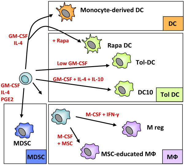
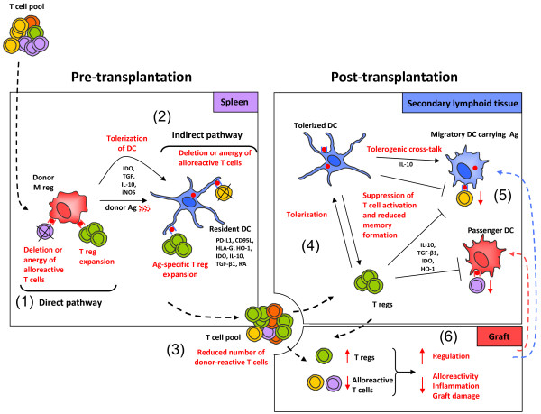
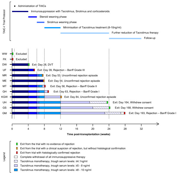
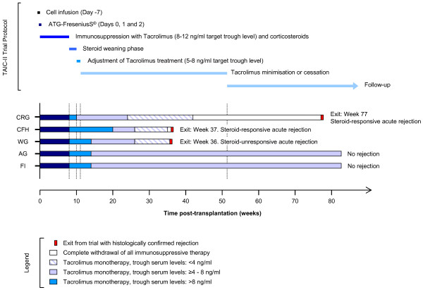
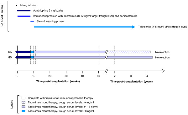
Several types of myeloid suppressor cell are currently being developed as cell-based immunosuppressive agents. Despite detailed knowledge about the molecular and cellular functions of these cell types, expert opinions differ on how to best implement such therapies in solid organ transplantation. Efforts in our laboratory to develop a cell-based medicinal product for promoting tolerance in renal transplant patients have focused on a type of suppressor macrophage, which we call the regulatory macrophage (M reg). Our favoured clinical strategy is to administer donor-derived M regs to recipients one week prior to transplantation. In contrast, many groups working with tolerogenic dendritic cells (DCs) advocate post-transplant administration of recipient-derived cells. A third alternative, using myeloid-derived suppressor cells, presumably demands that cells are given around the time of transplantation, so that they can infiltrate the graft to create a suppressive environment. On present evidence, it is not possible to say which cell type and treatment strategy might be clinically superior. This review seeks to position our basic scientific and early-stage clinical studies of human regulatory macrophages within the broader context of myeloid suppressor cell therapy in transplantation.


Figures

Figure 1
Figure 1
The spectrum of monocyte-derived suppressor APCs. Suppressor macrophages and DCs can be generated from monocytes using M-CSF or GM-CSF, with or without IL-4. Development of immature DCs into mature, activating DCs can be blocked by various substances, including rapamycin or dexamethasone and vitamin D. DCs can also be rendered tolerogenic by culture in low-dose GM-CSF or by addition of suppressive cytokines, such as IL-10 or TGF-β1. Mesenchymal stem cells (MSC) can induce a suppressor phenotype in co-cultured macrophages. Myeloid-derived suppressor cells (MDSC) can be generated by exposing monocytes/macrophages to tumour-secreted factors, most notably PGE2.
Figure 2
Figure 2
Proposed mode of M reg action. (1) When administered prior to transplantation, donor M regs migrate to spleen, where they present donor antigen through the direct pathway to alloreactive T cells and either delete or anergise them, or induce expansion of regulatory T cells. Human M regs have been shown to delete activated T cells through a contact-dependent mechanism and to suppress T cell proliferation through IDO; however, other suppressor mechanisms may also contribute to M reg function, such as IL-10 and TGF-β secretion, or iNOS activity. (2) It is likely that M regs serve as a source of donor alloantigen, which is captured and presented by immature recipient DCs to alloreactive T cells via the indirect and semi-direct pathways of alloantigen recognition. In consequence, responding T cells may be deleted or anergised, and antigen-specific T regs may be induced. (3) Through these mechanisms, the recipient T cell pool is enriched for T regs and depleted of donor-reactive T cells. (4) After transplantation, recipient T regs could induce tolerogenic DCs in secondary lymphoid organs. (5) Recipient tolerogenic DCs could then suppress activation of T cells. (6) In consequence, an immunological environment conducive to allograft acceptance is established.
Figure 3
Figure 3
Overview of the TAIC-I trial. Patients enrolled in the TAIC-I Study each received a kidney transplant from a deceased donor. The mean age of the patients was 46.3 years and 9/12 patients were male. The median HLA-A,-B and –DR mismatch was 5/6. Initially, patients were treated with a combination of tacrolimus (trough levels of 10–15 ng/ml), sirolimus (trough levels of 4–8 ng/ml) and corticosteroids. Cells were infused on day 5 post-transplant. Steroids were tapered in weeks 5 and 6. Sirolimus was withdrawn in weeks 7 and 8. If graft function remained stable, tacrolimus treatment was first minimised to trough tacrolimus levels of 8–10 ng/ml by week 12 and then to levels of 5–8 ng/ml by week 24. Further reductions in tacrolimus therapy were undertaken in patients with stable graft function and no histological evidence of rejection. Figure reproduced with permission from Hutchinson, JA. et al. Transplant International (2008) 21:728–741.
Figure 4
Figure 4
Overview of the TAIC-II trial. Patients enrolled in the TAIC-II Study each received a kidney transplant from a living donor. The mean age of the patients was 35.4 years and 4/5 patients were male. The median HLA-A,-B and –DR mismatch was 3/6. Cells were infused 5 days prior to transplantation. All patients received ATG induction therapy on days 0, 1 and 2. Initial maintenance immunosuppression comprised glucocorticoids and tacrolimus (8–12 ng/ml trough levels). Steroid therapy was withdrawn by week 10. Tacrolimus dosing was then adjusted into a target range of 5 – 8 ng/ml trough levels. From week 24 onwards, further reductions were made in tacrolimus monotherapy, leading to complete drug withdrawal in two patients.
Figure 5
Figure 5
Overview of the treatment of patients MM and CA. Both patients received a living-donor kidney transplant. M regs were infused 6 (MM) or 7 (CA) days prior to transplantation under cover of 2 mg/kg/day azathioprine. Initial maintenance immunosuppression comprised glucocorticoids and tacrolimus (>8 ng/ml trough levels). Steroid therapy was withdrawn by week 10. Tacrolimus dosing was then adjusted into a target range of 4 – 8 ng/ml trough levels.
Figure 6
Figure 6
Summary of a tacrolimus-minimisation study conducted by Shapiro et al.[[65]]. This clinical trial remains a benchmark study of minimised tacrolimus monotherapy in renal transplant recipients. 150 patients were treated with ATG induction therapy and bolus prednisone before being maintained on tacrolimus monotherapy. Over several months, tacrolimus was weaned in a stepwise fashion from 113 patients. The acute rejection rate prior to weaning was 37% and the acute rejection rate during weaning was 23%.

References

    1. Hutchinson JA, Riquelme P, Brem-Exner BG, Schulze M, Matthai M, Renders L, Kunzendorf U, Geissler EK, Fandrich F. Transplant acceptance-inducing cells as an immune-conditioning therapy in renal transplantation. Transpl Int. 2008;21:728–741. doi: 10.1111/j.1432-2277.2008.00680.x. - DOI - PubMed
    1. Hutchinson JA, Brem-Exner BG, Riquelme P, Roelen D, Schulze M, Ivens K, Grabensee B, Witzke O, Philipp T, Renders L. et al.A cell-based approach to the minimization of immunosuppression in renal transplantation. Transpl Int. 2008;21:742–754. doi: 10.1111/j.1432-2277.2008.00692.x. - DOI - PubMed
    1. Hutchinson JA, Riquelme P, Sawitzki B, Tomiuk S, Miqueu P, Zuhayra M, Oberg HH, Pascher A, Lutzen U, Janssen U. et al.Cutting Edge: immunological consequences and trafficking of human regulatory macrophages administered to renal transplant recipients. J Immunol. 2011;187:2072–2078. doi: 10.4049/jimmunol.1100762. - DOI - PubMed
    1. Harry RA, Anderson AE, Isaacs JD, Hilkens CM. Generation and characterisation of therapeutic tolerogenic dendritic cells for rheumatoid arthritis. Ann Rheum Dis. 2010;69:2042–2050. doi: 10.1136/ard.2009.126383. - DOI - PMC - PubMed
    1. Giannoukakis N, Phillips B, Finegold D, Harnaha J, Trucco M. Phase I (safety) study of autologous tolerogenic dendritic cells in type 1 diabetic patients. Diabetes Care. 2011;34:2026–2032. doi: 10.2337/dc11-0472. - DOI - PMC - PubMed

LinkOut - more resources