. 2013 Jan 28;24(2):159-68.
doi: 10.1016/j.devcel.2012.12.005.

A Rab10-dependent mechanism for polarized basement membrane secretion during organ morphogenesis

Affiliations

A Rab10-dependent mechanism for polarized basement membrane secretion during organ morphogenesis

David W Lerner et al. Dev Cell. .

Abstract

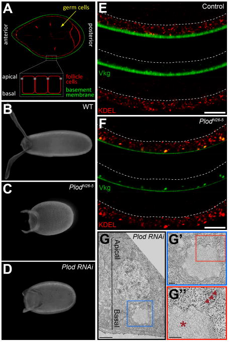
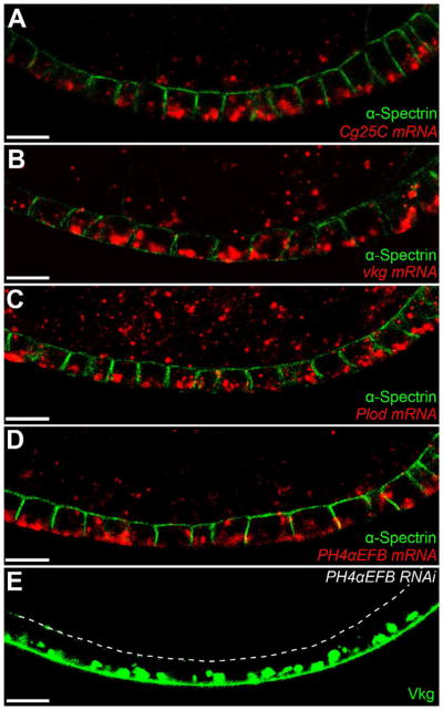
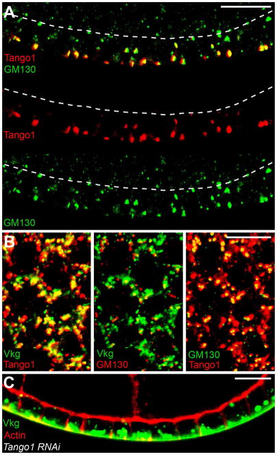
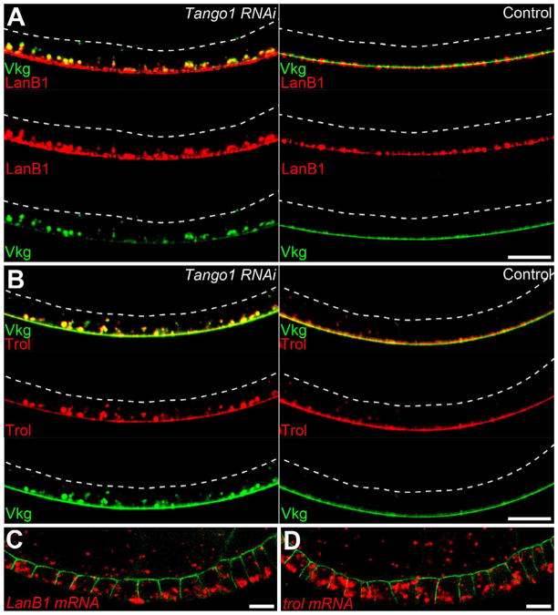
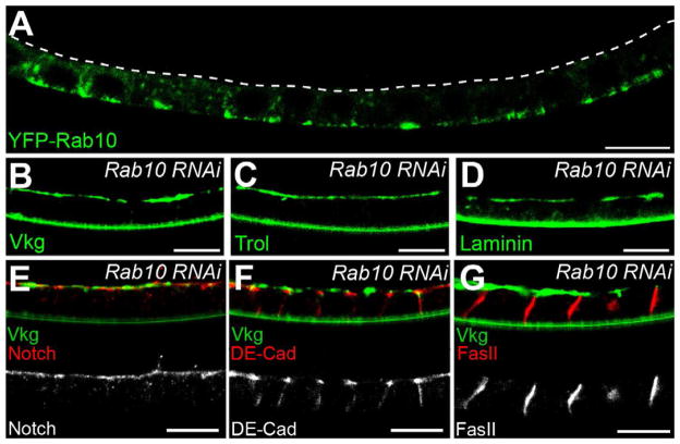
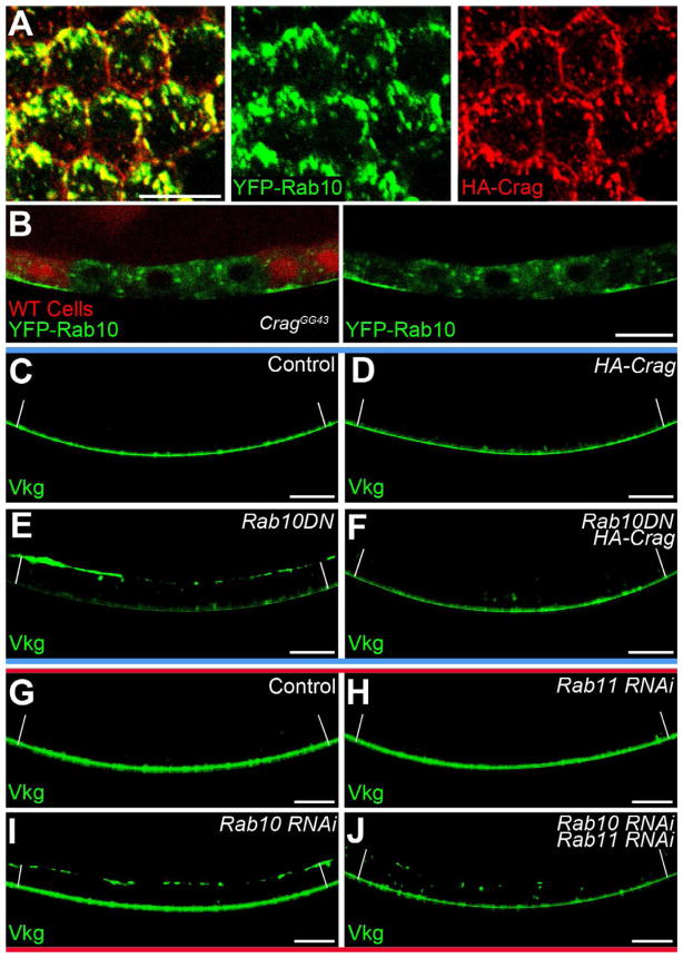
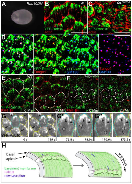
Basement membranes (BMs) are specialized extracellular matrices that are essential for epithelial structure and morphogenesis. However, little is known about how BM proteins are delivered to the basal cell surface or how this process is regulated during development. Here, we identify a mechanism for polarized BM secretion in the Drosophila follicle cells. BM proteins are synthesized in a basal endoplasmic reticulum (ER) compartment from localized mRNAs and are then exported through Tango1-positive ER exit sites to basal Golgi clusters. Next, Crag targets Rab10 to structures in the basal cytoplasm, where it restricts protein delivery to the basal surface. These events occur during egg chamber elongation, a morphogenetic process that depends on follicle cell planar polarity and BM remodeling. Significantly, Tango1 and Rab10 are also planar polarized at the basal epithelial surface. We propose that the spatial control of BM production along two tissue axes promotes exocytic efficiency, BM remodeling, and organ morphogenesis.


Figures

Figure 1
Figure 1. PlodN26–5 disrupts the secretion of Col IV
(A) Overview of egg chamber structure at stage 8. Actin (red), Vkg-GFP (green). (B–D) Representative egg shapes for (B) wild type, (C) PlodN26-5, and (D) Plod-RNAi. (E) In wild type, proteins with the KDEL ER retention signal are throughout the cell, while Vkg-GFP is in the BM. (F) In PlodN26-5, Vkg-GFP punctae overlap with KDEL near the basal surface, indicating ER accumulation. (E and F) Dashed lines mark the apical surface, and scale bars are 10 μm. (G) TEM of a single Plod-RNAi follicle cell showing a distended ER region in the basal cytoplasm. Scale bar is 1 μm. (G′ and G″) Magnifications of the image in (G). Ribosomes decorate the distended ER cisterna (asterisk) and adjacent, normal ER membranes (arrowheads). Scale bars are 200 nm. (E–G) Experiments performed at stages 7–8. See also Figure S1.
Figure 2
Figure 2. Col IV is synthesized in the basal ER
(A–E) mRNAs encoding the Col IV α-chains (A) Cg25C and (B) Vkg, and the Col IV biosynthetic enzymes (C) Plod and (D) PH4αEFB are all enriched in the basal cytoplasm. α-Spectrin marks the apical and lateral epithelial surfaces. (E) PH4αEFB-RNAi causes Vkg-GFP to accumulate in the basal ER. The dashed line marks the apical surface. Experiments performed at stages 7–8. Scale bars are 10 μm. See also Figure S2.
Figure 3
Figure 3. Tango1 mediates Col IV ER exit predominantly at basal tER sites
(A) Tango1 is strongly enriched at basal tER-Golgi units, as indicated by the cis-Golgi marker, GM130. Dashed lines mark the apical surface. (B) An optical section through the basal cytoplasm showing co-localization between Tango1, GM130 and Vkg-GFP. (C) Tango1-RNAi causes Vkg-GFP to accumulate in the basal ER. Experiments performed at stages 7–8. Scale bars are 10 μm. See also Figure S3.
Figure 4
Figure 4. Laminin and Trol are also enriched in the basal cytoplasm
(A and B) Tango1-RNAi causes (A) LanB1 and (B) Trol to accumulate in the basal ER with Vkg-GFP. Dashed lines mark the apical surface. (C) LanB1 mRNA is basally enriched. (D) trol mRNA does not show a basal bias. (C and D) α-Spectrin marks the apical and lateral epithelial surfaces. Experiments performed at stages 7–8. Scale bars are 10 μm. See also Figure S4.
Figure 5
Figure 5. Rab10 promotes polarized BM secretion
(A) YFP-Rab10 is enriched near the basal follicle cell surface. The dashed line marks the apical surface. (B–D) Rab10-RNAi mis-targets (B) Col IV (Vkg-GFP), (C) Perlecan (Trol-GFP) and (D) Laminin to the apical surface. (E-G) Rab10-RNAi does not affect the localization of (E) apical (Notch), (F) junctional (DE-Cadherin), or (G) lateral (FasII) trans-membrane proteins. Rab10 knockdown is shown by apical Vkg-GFP. Experiments performed at stages 7–8, except (C and D), which are stage 9. Scale bars are 10 μm. See also Figure S5.
Figure 6
Figure 6. Crag regulates Rab10 during polarized BM secretion
(A) Optical section near the basal surface showing YFP-Rab10 and HA-Crag co-localization. (B) A CragGG43 follicle cell clone shows a redistribution of YFP-Rab10 away from the basal surface. (C–F) Interaction between UAS-HA-Crag and UAS-Rab10.T23N. (C–D) Vkg-GFP localization is normal in (C) wild-type and (D) UAS-HA-Crag follicle cells. (E) A UAS-Rab10.T23N dominant negative transgene causes Vkg-GFP to accumulate on the apical surface. (F) Co-expression of UAS-HA-Crag with UAS-Rab10.T23N suppresses this phenotype. (E–F) A YFP on Rab10.T23N contributes to the fluorescent signal in the cytoplasm. (G–J) Interaction between Rab10-RNAi and Rab11-RNAi. (G and H) Vkg-GFP localization is normal in (G) wild-type and (H) Rab11-depleted cells. (I) Rab10 depletion causes Vkg-GFP to accumulate on the apical surface. (J) Co-depletion of Rab10 and Rab11 eliminates the apical Vkg-GFP and increases the cytoplasmic signal. (C–J) White lines extend between the apical and basal epithelial surfaces. Experiments performed at stages 7–8. Scale bars are 10 μm. See also Figure S6.
Figure 7
Figure 7. The BM exocytic machinery is planar polarized at the basal epithelial surface
(A) Rab10.T23N expression produces round eggs. (B) YFP-Rab10 is planar polarized at the level of the basal actin filaments. (C) YFP-Rab10’s planar polarization is lost in fat2N103-2 epithelia. (D) Tango1 and GM130 are also planar polarized, and partially overlap with YFP-Rab10. (E) Live imaging reveals that YFP-Rab10 is enriched at the trailing edge of each migrating cell. (F) fat2N103–2 epithelia fail to migrate and YFP-Rab10 is mis-polarized. (B–F) Scale bars are 10 μm. (E and F) FM4–64 marks cell membranes. Dashed lines mark the same 3–4 cells at each time point. (G) TIRF microscopy reveals that larger Rab10-positive structures are largely immobile (arrows), while smaller Rab10-positive structures (circles in G′ and G″) move rapidly through the cytoplasm. Scale bars are 2 μm. (H) Speculative model for how planar polarization of BM secretion would synergize with follicle cell migration to create the unusual BM fibrils associated with egg chamber elongation. Illustration adapted from (Bilder and Haigo, 2012). Experiments performed at stages 7–8. See also Figure S7 and Movies S1–S3.

References

    1. Arnaoutova I, George J, Kleinman HK, Benton G. Basement membrane matrix (BME) has multiple uses with stem cells. Stem Cell Rev. 2012;8:163–169. - PubMed
    1. Babbey CM, Ahktar N, Wang E, Chen CC, Grant BD, Dunn KW. Rab10 regulates membrane transport through early endosomes of polarized Madin-Darby canine kidney cells. Mol Biol Cell. 2006;17:3156–3175. - PMC - PubMed
    1. Babbey CM, Bacallao RL, Dunn KW. Rab10 associates with primary cilia and the exocyst complex in renal epithelial cells. Am J Physiol Renal Physiol. 2010;299:F495–506. - PMC - PubMed
    1. Bilder D, Haigo SL. Expanding the morphogenetic repertoire: perspectives from the Drosophila egg. Dev Cell. 2012;22:12–23. - PMC - PubMed
    1. Boll W, Partin JS, Katz AI, Caplan MJ, Jamieson JD. Distinct pathways for basolateral targeting of membrane and secretory proteins in polarized epithelial cells. Proc Natl Acad Sci U S A. 1991;88:8592–8596. - PMC - PubMed

Publication types

MeSH terms

Substances