. 2013 May-Jun;7(3):221-8.
doi: 10.4161/pri.23807. Epub 2013 Jan 31.

Beta conformation of polyglutamine track revealed by a crystal structure of Huntingtin N-terminal region with insertion of three histidine residues

Affiliations

Beta conformation of polyglutamine track revealed by a crystal structure of Huntingtin N-terminal region with insertion of three histidine residues

Meewhi Kim. Prion. 2013 May-Jun.

Abstract

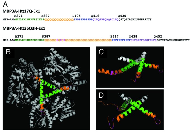
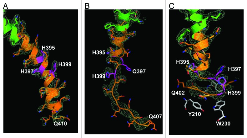
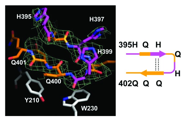
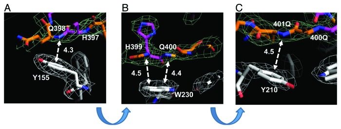
Huntington disease is an autosomal-dominant neurodegenerative disorder caused by a polyglutamine (polyQ) expansion (> 35Q) in the first exon (EX1) of huntingtin protein (Htt). mHtt protein is thought to adopt one or more toxic conformation(s) that are involved in pathogenic interactions in cells . However, the structure of mHtt is not known. Here, we present a near atomic resolution structure of mHtt36Q-EX1. To facilitate crystallization, three histidine residues (3H) were introduced within the Htt36Q stretch resulting in the sequence of Q 7HQHQHQ 27. The Htt36Q3H region adopts α-helix, loop, β-hairpin conformations. Furthermore, we observed interactions between the backbone of the Htt36Q3H β-strand with the aromatic residues mimicking putative-toxic interactions with other proteins. Our findings support previous predictions that the expanded mHtt-polyQ region adopts a β-sheet structure. Detailed structural information about mHtt improves our understanding of the pathogenic mechanisms in HD and other polyQ expansion disorders and may form the basis for rational design of small molecules that target toxic conformations of disease-causing proteins.

Keywords: Huntingtin; amyloid; crystallography; hairpin; polyglutamine; prion; sggregation; staxia; β-strand; β-turn.


Figures

None
Figure 1. The structure of MBP-Htt36Q3H-EX1. (A) Amino acid sequence of MBP-Htt17Q-EX1 and MBP-Htt36Q3H-EX1 expression constructs. MBP3A denotes the maltose binding protein followed by a three alanines linker. In both constructs position M371 corresponds to M1 in Htt sequence. The sequence of Htt-Ex1 is composed of N-terminal N17 region (green), polyQ region (orange), His (pink), PolyP region (blue) and mixed P/Q region (purple). Three His residues (pink) are inserted within poly36Q stretch in MBP-Htt36Q3H-EX1 construct. In both constructs identical 19 amino acids C-terminal tag was added to facilitate crystallization (black).(B) The trimer of MBP-Htt36Q3H-EX1. (C, D) Htt36Q3H-EX1 trimers in the crystal X1 (C) and X2 (D). On panels (B), (C) and (D) the MBP protein (gray), 3A linker (gray), Htt-N17 (green) and Htt-polyQ (orange) regions are shown for each of the three MBP-Htt36Q3H-EX1 molecules (A, B, C) in the asymmetric unit of the crystal.
None
Figure 2. The Htt36Q3H region in α-helical, loop and β-hairpin conformations. (A) The α-helical structure of Htt36Q3H (Q388-Q410) (C1 molecule from crystal X1). (B) The α-helical structure of Htt36Q3H (Q388-H395) followed by a loop (Q396-Q407) (C1 molecule from crystal X2). (C) The α-helical structure of Htt36Q3H (Q388-Q394) followed by a β-hairpin (H395-Q401) (C2 molecule from crystal X1). On (A), (B) and (C) Htt-N17 (green), Htt-polyQ (orange) and His residues (pink) are labeled and numbered on the ribbon diagram. The supportive electron density maps (2Fo-Fc) at 1σ are shown by a green mesh.
None
Figure 3. The Htt36Q3H region in β-hairpin conformation. The hairpin structure of Htt36Q3H (H395-Q401) (C1 molecule from crystal X1). The Gln (orange) and His (pink) residues are labeled and numbered on the stick diagram. The hydrogen bonds are shown by a dotted line. The Y210 and W230 proximal residues from MBP are also shown. The supportive electron density maps (2Fo-Fc) at 1σ are shown by green mesh for Htt36Q3H. The diagram of structure is depicted in a schematic form on the right panel.
None
Figure 4. Intermolecular interactions between β−hairpin of Htt36Q3H and aromatic residues in MBP. (A) The interaction between the amide backbone of Q398 in Htt36Q3H and Y155 in MBP. (B) The interactions between the amide backbones of H399 and Q400 in Htt36Q3H and W230 in MBP. (C) The interactions between the amide backbone of Q401 in Htt36Q3H and Y210 in MBP. On panels (A), (B) and (C) the interactions at different angles are shown for C1 molecule in crystal X1. The Gln (orange) and His (pink) residues are labeled on the stick diagram The interacting residues are numbered and the interactions are shown with a dotted arrow line with interaction distances indicated in angstroms (Å). The interaction distances were measured from the backbone of the residues in the Htt36Q3H β-strand to the center of the aromatic ring residues in MBP. The supportive electron density maps (2Fo-Fc) at 1σ are shown by a green mesh for Htt38Q3H and a white mesh for MBP.

References

    1. Vonsattel JP, DiFiglia M. Huntington disease. J Neuropathol Exp Neurol. 1998;57:369–84. doi: 10.1097/00005072-199805000-00001. - DOI - PubMed
    1. The Huntington's Disease Collaborative Research Group A novel gene containing a trinucleotide repeat that is expanded and unstable on Huntington disease chromosomes. Cell. 1993;72:971–83. doi: 10.1016/0092-8674(93)90585-E. - DOI - PubMed
    1. Tobin AJ, Signer ER. Huntington’s disease: the challenge for cell biologists. Trends Cell Biol. 2000;10:531–6. doi: 10.1016/S0962-8924(00)01853-5. - DOI - PubMed
    1. Ross CA. Polyglutamine pathogenesis: emergence of unifying mechanisms for Huntington’s disease and related disorders. Neuron. 2002;35:819–22. doi: 10.1016/S0896-6273(02)00872-3. - DOI - PubMed
    1. Rubinsztein DC. Lessons from animal models of Huntington’s disease. Trends Genet. 2002;18:202–9. doi: 10.1016/S0168-9525(01)02625-7. - DOI - PubMed

Publication types