

Randomized clinical trial evaluating intravitreal ranibizumab or saline for vitreous hemorrhage from proliferative diabetic retinopathy
- PMID: 23370902
- PMCID: PMC4217122
- DOI: 10.1001/jamaophthalmol.2013.2015
Randomized clinical trial evaluating intravitreal ranibizumab or saline for vitreous hemorrhage from proliferative diabetic retinopathy
Abstract
Importance: Vascular endothelial growth factor plays a role in proliferative diabetic retinopathy (PDR). Intravitreal injection of saline has been shown potentially to lead to improved visual acuity compared with observation alone in eyes with vitreous hemorrhage. Therefore, it is important to determine if intravitreal anti-vascular endothelial growth factor can reduce vitrectomy rates (and risks associated with vitrectomy) compared with saline for vitreous hemorrhage from PDR that precludes placement or confirmation of complete panretinal photocoagulation.
Objective: To evaluate intravitreal ranibizumab compared with intravitreal saline injections on vitrectomy rates for vitreous hemorrhage from PDR.
Design: Phase 3, double-masked, randomized, multicenter clinical trial. Data reported were collected from June 2010 to March 2012 and include 16 weeks of follow-up.
Setting: Community-based and academic-based ophthalmology practices specializing in retinal diseases.
Participants: Two hundred sixty-one eyes of 261 study participants, who were at least 18 years of age with type 1 or type 2 diabetes mellitus. Study eyes had vitreous hemorrhage from PDR precluding panretinal photocoagulation completion.
Intervention: Eyes were randomly assigned to 0.5-mg intravitreal ranibizumab (n = 125) or intravitreal saline (n = 136) at baseline and 4 and 8 weeks.
Main outcome measure: Cumulative probability of vitrectomy within 16 weeks.
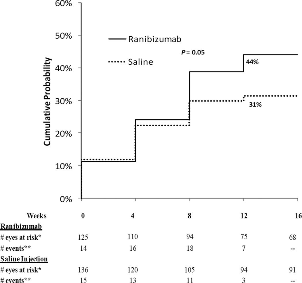
Results: Cumulative probability of vitrectomy by 16 weeks was 12% with ranibizumab vs 17% with saline (difference, 4%; 95% CI, -4% to 13%) and of complete panretinal photocoagulation without vitrectomy by 16 weeks was 44% and 31%, respectively (P = .05). The mean (SD) visual acuity improvement from baseline to 12 weeks was 22 (23) letters and 16 (31) letters, respectively (P = .04). Recurrent vitreous hemorrhage occurred within 16 weeks in 6% and 17%, respectively (P = .01). One eye developed endophthalmitis after saline injection.
Conclusions and relevance: Overall, the 16-week vitrectomy rates were lower than expected in both groups. This study suggests little likelihood of a clinically important difference between ranibizumab and saline on the rate of vitrectomy by 16 weeks in eyes with vitreous hemorrhage from PDR. Short-term secondary outcomes including visual acuity improvement, increased panretinal photocoagulation completion rates, and reduced recurrent vitreous hemorrhage rates suggest biologic activity of ranibizumab. Long-term benefits remain unknown. Whether vitrectomy rates after saline or ranibizumab injection are different than observation alone cannot be determined from this study.
Trial registration: The study is listed on www.clinicaltrials.gov, under identifier NCT00996437 (website registration date October 14, 2009).
Figures

References
-
- Kempen JH, O`Colmain BJ, Leske MC, et al. The prevalence of diabetic retinopathy among adults in the United States. Arch Ophthalmol. 2004;122:552–563. - PubMed
-
- Flynn HJ, Chew E, Simons B, et al. Pars plana vitrectomy in the early treatment diabetic retinopathy study: ETDRS report number 17. Ophthalmology. 1992;99:1351–1357. - PubMed
-
- American Society of Retina Specialist. PAT Survey. 2008
-
- Smiddy WE, Flynn HW., Jr Vitrectomy in the management of diabetic retinopathy. Surv Ophthalmol. 1999;43:491–507. - PubMed
-
- Yanoff M, Duker JS. Ophthalmology. 3rd ed. Vitrectomy: Mosby Elsevier; 1999.
Publication types
MeSH terms
Substances
Associated data
Grants and funding
LinkOut - more resources
Full Text Sources
Other Literature Sources
Medical
