. 2013 May;48(5):665-73.
doi: 10.1165/rcmb.2011-0273OC.

Smoke extract impairs adenosine wound healing: implications of smoke-generated reactive oxygen species

Affiliations

Smoke extract impairs adenosine wound healing: implications of smoke-generated reactive oxygen species

Diane S Allen-Gipson et al. Am J Respir Cell Mol Biol. 2013 May.

Abstract

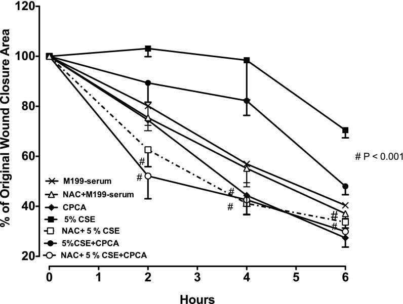
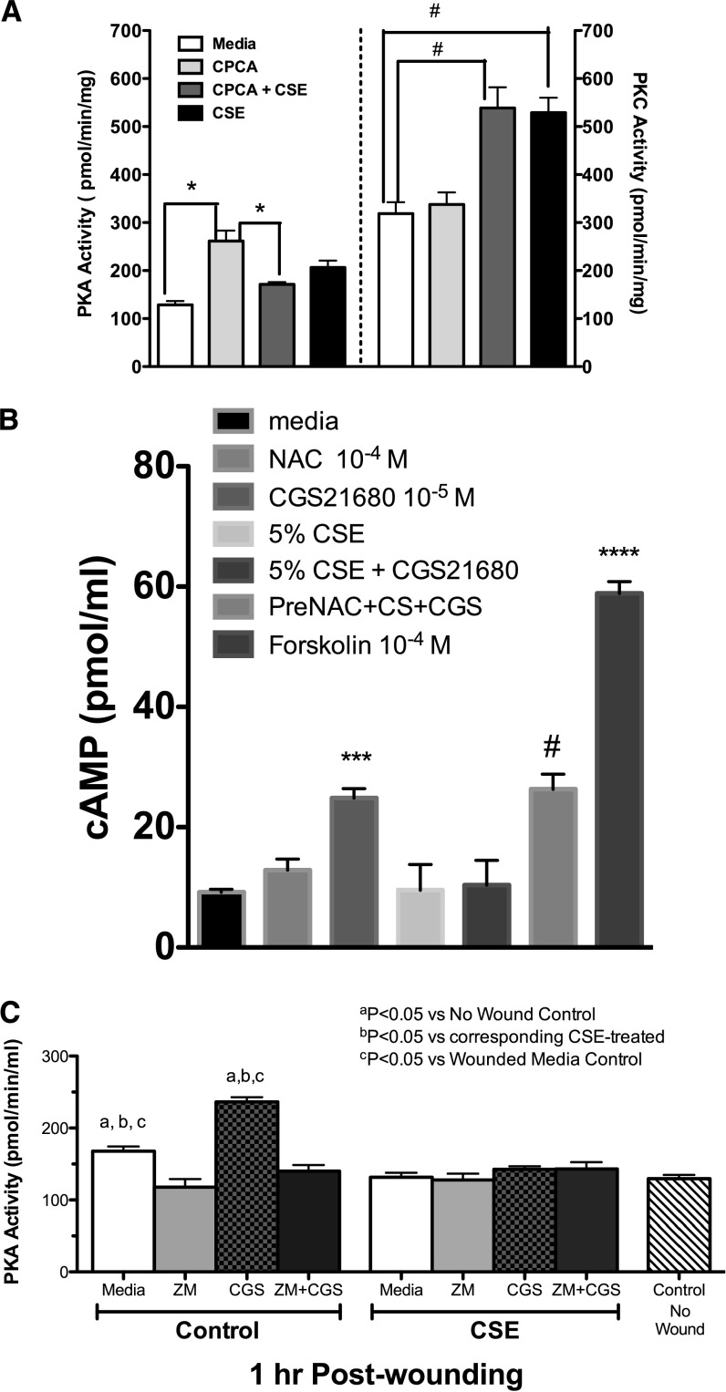
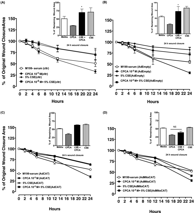
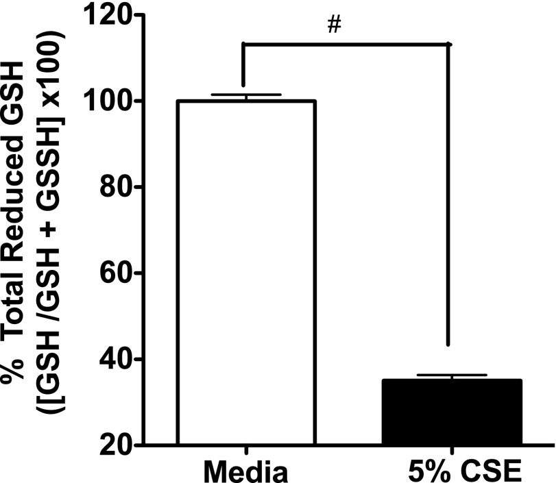
Adenosine concentrations are elevated in the lungs of patients with asthma and chronic obstructive pulmonary disease, where it balances between tissue repair and excessive airway remodeling. We previously demonstrated that the activation of the adenosine A2A receptor promotes epithelial wound closure. However, the mechanism by which adenosine-mediated wound healing occurs after cigarette smoke exposure has not been investigated. The present study investigates whether cigarette smoke exposure alters adenosine-mediated reparative properties via its ability to induce a shift in the oxidant/antioxidant balance. Using an in vitro wounding model, bronchial epithelial cells were exposed to 5% cigarette smoke extract, were wounded, and were then stimulated with either 10 μM adenosine or the specific A2A receptor agonist, 5'-(N-cyclopropyl)-carboxamido-adenosine (CPCA; 10 μM), and assessed for wound closure. In a subset of experiments, bronchial epithelial cells were infected with adenovirus vectors encoding human superoxide dismutase and/or catalase or control vector. In the presence of 5% smoke extract, significant delay was evident in both adenosine-mediated and CPCA-mediated wound closure. However, cells pretreated with N-acetylcysteine (NAC), a nonspecific antioxidant, reversed smoke extract-mediated inhibition. We found that cells overexpressing mitochondrial catalase repealed the smoke extract inhibition of CPCA-stimulated wound closure, whereas superoxide dismutase overexpression exerted no effect. Kinase experiments revealed that smoke extract significantly reduced the A2A-mediated activation of cyclic adenosine monophosphate-dependent protein kinase. However, pretreatment with NAC reversed this effect. In conclusion, our data suggest that cigarette smoke exposure impairs A2A-stimulated wound repair via a reactive oxygen species-dependent mechanism, thereby providing a better understanding of adenosine signaling that may direct the development of pharmacological tools for the treatment of chronic inflammatory lung disorders.


Figures

<i>Figure 1.</i>
Figure 1.
N-acetylcysteine (NAC) attenuates the smoke extract–mediated inhibition of adenosine A2A–stimulated wound closure. Bovine bronchial epithelial cells (BBECs) were pretreated overnight with 100 μM of NAC, a potent antioxidant. After pretreatment, the cells were wounded and then stimulated with or without 10 μM 5′-(N-cyclopropyl)–carboxamido–adenosine (CPCA) ± 5% cigarette smoke extract (CSE) in serum-free media. Cells pretreated with NAC significantly attenuated the smoke extract–mediated inhibition of adenosine-stimulated wound closure. Data represent the mean ± SE of triplicate wells within a single experiment. #P < 0.001, in comparison with smoke extract–exposed group and/or CPCA + smoke extract–exposed group at the same time point, according to ANOVA. The experiment was repeated twice using different preparations of BBECs, with similar results.
<i>Figure 2.</i>
Figure 2.
NAC reversed the smoke extract–mediated inhibition of adenosine A2A receptor (A2AAR)–stimulated cyclic adenosine monophosphate (cAMP)–dependent protein kinase (PKA) activation in wounded bovine bronchial epithelial cells. BBECs were pretreated overnight with 100 μM NAC. The next day, cells were incubated in serum-free media, wounded, stimulated with or without 10 μM CPCA ± 5% smoke extract, and assessed for kinase activity. Smoke extract noticeably (**P < 0.01) reduced the CPCA-mediated activation of PKA, compared with the CPCA-stimulated group. NAC significantly (#P < 0.001) reversed the smoke extract–mediated inhibition of adenosine-stimulated PKA activation, compared with the smoke extract + CPCA − NAC–exposed group, according to ANOVA. Data represent the mean ± SE of triplicate wells within a single experiment. The experiment was repeated twice using different preparations of BBECs, with similar results.
<i>Figure 3.</i>
Figure 3.
Smoke extract exposure blunts the A2A-mediated activation of cAMP and cAMP-dependent kinase (PKA), and stimulates protein kinase C (PKC) activation in wounded bronchial epithelial cells. (A) Direct kinase activity measurement was used to determine the effects of smoke extract on the CPCA-mediated activation of PKA (left y axis) and the activation by smoke extract of PKC (right y axis). *P < 0.01; #P < 0.001. (B) CSE exposure blunts the A2A-mediated activation of cAMP. Monolayers of Nuli-1 cells were either pretreated for 1 hour with NAC (100 nM) and stimulated with CGS21680 (CGS; an A2A agonist) ± 5% smoke extract (CS), and cAMP activity was assayed. CGS21680 significantly increased the activation of cAMP, compared with media control cells (***P < 0.001). Smoke extract noticeably reduced the CGS21680-mediated activation of cAMP, compared with the CGS21680-stimulated group. NAC significantly (#P < 0.001) reversed the smoke extract–mediated inhibition of adenosine-stimulated cAMP activation, compared with the smoke extract + CGS21680 − NAC–exposed group. As a positive control, some cells were treated with 100 nM forskolin (a cAMP activator; ****P < 0.0001). Data represent the mean ± SE of triplicate wells within a single experiment. (C) Blocking A2A receptor reduces PKA activation in wounded cells. Wounded Nuli-1 cells were either pretreated for 1 hour with ZM241385 (ZM; 100 nM) and stimulated with CGS21680 ± 5% smoke extract, and PKA was assayed. CGS21680 significantly increased the activation of cAMP, compared with media control cells (P < 0.05). Smoke extract notably blunted CGS21680-stimulated PKA. Data represent the mean ± SE of three separate experiments performed in triplicate.
<i>Figure 4.</i>
Figure 4.
Smoke extract exposure generates hydrogen peroxide (H2O2) in wounded bronchial epithelial cells. Wounded monolayers of BEAS-2B cells were exposed to 5% smoke extract, and after 0, 2, 4, 6, and 24 hours, the supernatants were collected, and H2O2 concentrations were measured. A significant release of H2O2 occurred in a time-dependent fashion (**P < 0.01, compared with baseline; #P < 0.001, compared with baseline). Error bars represent the SE of three separate experiments performed in duplicate. RFU, relative fluorescence units.
<i>Figure 5.</i>
Figure 5.
The adenovirus-mediated expression of mitochondrially targeted catalase blocks the smoke extract–induced inhibition of adenosine-stimulated wound closure. Smoke extract blocks CPCA-mediated wound closure in (A) noninfected (cltr), (B) empty vector (AdEmpty)–infected, and (C) adenovirus encoding H2O2 scavenging enzyme catalase (AdCat)–infected BEAS-2B cells in serum-free media, compared with CPCA alone (*P < 0.05). (D) In contrast, mitochondrially targeted catalase (AdMitoCat) reversed the CSE inhibition of CPCA-mediated wound closure, compared with CPCA alone (*P > 0.05). Error bars represent the SE of three separate experiments performed in duplicate. NS, no significance.
<i>Figure 6.</i>
Figure 6.
Smoke extract reduces glutathione (GSH) concentrations in bronchial epithelial cells. Confluent monolayers of BEAS-2B cells were exposed to 5% smoke extract for 1 hour, and total GSH concentrations were measured as described in Materials and Methods. Data represent the mean ± SE of triplicate wells within a single experiment. The experiment was repeated twice with similar results. #P < 0.01, versus media control cells according to ANOVA.
<i>Figure 7.</i>
Figure 7.
Smoke extract exposure increases intracellular adenosine in bronchial epithelial cells. The determination of adenosine using isocratic, reversed-phase, high-performance liquid chromatography, coupled with an online detector for ultraviolet light absorbance and electrochemical activity, was performed in BEAS-2B. Cells were treated with either 5% smoke extract or medium for 1 hour. The smoke extract–treated cells demonstrated a significant elevation of intracellular adenosine (85 ± 10.7 pmole/106 cells; #P < 0.01, compared with 39 ± 11.7 pmole/106 control cells). Data represent the mean ± SD of two separate experiments performed in duplicate.

References

    1. Eckle T, Koeppen M, Eltzschig HK. Role of extracellular adenosine in acute lung injury. Physiology (Bethesda) 2009;24:298–306. - PubMed
    1. Zhou Y, Murthy JN, Zeng D, Belardinelli L, Blackburn MR. Alterations in adenosine metabolism and signaling in patients with chronic obstructive pulmonary disease and idiopathic pulmonary fibrosis. PLoS ONE. 2010;5:e9224. - PMC - PubMed
    1. Fredholm BB, Jzerman I AP, Jacobson KA, Klotz KN, Linden J. International Union of Pharmacology: XXV. Nomenclature and classification of adenosine receptors. Pharmacol Rev. 2001;53:527–552. - PMC - PubMed
    1. Linden J. Molecular approach to adenosine receptors: receptor-mediated mechanisms of tissue protection. Annu Rev Pharmacol Toxicol. 2001;41:775–787. - PubMed
    1. Blackburn MR. Too much of a good thing: adenosine overload in adenosine–deaminase–deficient mice. Trends Pharmacol Sci. 2003;24:66–70. - PubMed

Publication types

MeSH terms

LinkOut - more resources