Review
. 2013 Feb 1;112(3):549-61.
doi: 10.1161/CIRCRESAHA.111.249243.

Immunogenicity of pluripotent stem cells and their derivatives

Affiliations
Review

Immunogenicity of pluripotent stem cells and their derivatives

Patricia E de Almeida et al. Circ Res. .

Abstract

The ability of pluripotent stem cells to self-renew and differentiate into all somatic cell types brings great prospects to regenerative medicine and human health. However, before clinical applications, much translational research is necessary to ensure that their therapeutic progenies are functional and nontumorigenic, that they are stable and do not dedifferentiate, and that they do not elicit immune responses that could threaten their survival in vivo. For this, an in-depth understanding of their biology, genetic, and epigenetic make-up and of their antigenic repertoire is critical for predicting their immunogenicity and for developing strategies needed to assure successful long-term engraftment. Recently, the expectation that reprogrammed somatic cells would provide an autologous cell therapy for personalized medicine has been questioned. Induced pluripotent stem cells display several genetic and epigenetic abnormalities that could promote tumorigenicity and immunogenicity in vivo. Understanding the persistence and effects of these abnormalities in induced pluripotent stem cell derivatives is critical to allow clinicians to predict graft fate after transplantation, and to take requisite measures to prevent immune rejection. With clinical trials of pluripotent stem cell therapy on the horizon, the importance of understanding immunologic barriers and devising safe, effective strategies to bypass them is further underscored. This approach to overcome immunologic barriers to stem cell therapy can take advantage of the validated knowledge acquired from decades of hematopoietic stem cell transplantation.


Figures

Figure 1
Figure 1
Percentage of Asian (A, n=797), Black (B, n=441), and White (W, n=5087) patients HLA matched using ten cohorts of 150 cadaveric organ donors. HLA mismatch grades was based on criteria used for allocation of cadaveric kidney donors in the UK: 1) zero HLA-A, HLA-B, and HLA-DR mismatch (0.0.0); 2) zero HLA-DR mismatch with no more than a single HLA-A or HLA-B mismatch (1.0.0 or 0.1.0); 3) zero HLA-DR mismatch with no more than a single HLA-A and a single HLA-B mismatch (1.1.0); 4) zero HLA-DR mismatch (*.*. 0). Reprint with permission.
Figure 2
Figure 2
Mechanisms for generating minor histocompatibility antigens in pluripotent stem cells. Polymorphisms induced in ES and iPS cells can result in expression of proteins and peptides that are distinct from those in the donor cells. Upon proteolytic degradation, these peptides are transported by the peptide transporter into the endoplasmic reticulum (ER), where they can bind to HLA molecules and pass through the Golgi apparatus to be presented at the cell surface as a complex with HLA and be recognized as foreign by donor T cells.
Figure 3
Figure 3
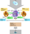
Simplified schema exemplifying an immune response to a stem cell-derived cellular therapeutic. Dendritic cells acquire antigens from the graft for presentation to T cells and NK cells, which mount specific responses following antigen receptor activation (Signal 1). Upon TCR–MHC interactions, co-stimulation (Signal 2) and pro-inflammatory cytokines (Signal 3), such as interleukin-12 (IL-12) and type I IFNs, can promote the activation and clonal expansion of T cells. Similarly, resting NK cells may also receive signals via activating receptors (Signal 2) and pro-inflammatory cytokine receptors (Signal 3). Activated T cells and NK cells can generate cytotoxic responses against the graft, resulting in rejection. Figure adapted from Sun & Lanier (2011).
Figure 4
Figure 4
Mean fluorescence intensity of various HLA proteins in various undifferentiated and differentiated human ES cell lines. The expression of HLA class I, HLA class II (HLA-DP,-DQ,-DR), and the non-classical HLA-I HLA-G was determined in two undifferentiated human ES cell lines (H9 and H13), embryonic bodies from in vitro differentiated human ES cells, in vivo differentiated human ES cells-teratomas; cervix epithelial cell line (HeLa). Dashed lines represent background control staining and solid lines demonstrate expression of specific antigens. Median fluorescence intensity staining is indicated at the top of each box. Reprint with permission.
Figure 5
Figure 5
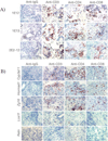
Extensive infiltration of T cells in regressing teratomas formed by syngeneic iPS and transgenic ES cells. A) T-cell infiltration in teratomas formed by syngeneic episomal-derived iPS cells from two different passages (1E-12, 1E-13) and after LoxP/Cre-mediated deletion of the reprogramming factor expression cassette from the integrated copy of episomal vector (2E2-12). B) Ectopic expression of Cyp3a11, Hormad1, and Zg16 in syngeneic mouse ES cells elicited infiltration of T cells in the teratomas. Few infiltrating T cells were detectable in the teratomas formed by Lce1f- and Retn-B6-expressing mouse ES cells. Reprint with permission.
Figure 6
Figure 6
Longitudinal bioluminescence imaging of human ES cells implanted intramuscularly in mice demonstrating that triple-costimulatory blockade therapy (COSTIM) administered at days 0, 2, 4, and 6 prevented rejection of human ES cells. COSTIM refers to a combination of CTLA4-Ig, anti-LFA-1, and anti-CD40L. COSTIM was remarkably more efficient than monotherapy with anti-LFA-1, CTLA4-Ig, or anti-CD40L. Reprint with permission.

Similar articles

Cited by

References

    1. Thomson JA, Itskovitz-Eldor J, Shapiro SS, Waknitz MA, Swiergiel JJ, Marshall VS, Jones JM. Embryonic stem cell lines derived from human blastocysts. Science. 1998;282:1145–1147. - PubMed
    1. Reubinoff BE, Pera MF, Fong C-Y, Trounson A, Bongso A. Embryonic stem cell lines from human blastocysts: somatic differentiation in vitro. Nat Biotech. 2000;18:399–404. - PubMed
    1. Soundararajan P, Lindsey BW, Leopold C, Rafuse VF. Easy and rapid differentiation of embryonic stem cells into functional motoneurons using sonic hedgehog-producing cells. Stem Cells. 2007;25:1697–1706. - PubMed
    1. Schroeder IS, Rolletschek A, Blyszczuk P, Kania G, Wobus AM. Differentiation of mouse embryonic stem cells to insulin-producing cells. Nat Protoc. 2006;1:495–507. - PubMed
    1. Barberi T, Klivenyi P, Calingasan NY, Lee H, Kawamata H, Loonam K, Perrier AL, Bruses J, Rubio ME, Topf N, Tabar V, Harrison NL, Beal MF, Moore MA, Studer L. Neural subtype specification of fertilization and nuclear transfer embryonic stem cells and application in parkinsonian mice. Nat Biotechnol. 2003;21:1200–1207. - PubMed

Publication types

MeSH terms

LinkOut - more resources