

A quantitative study of fixation stability in amblyopia
- PMID: 23372053
- PMCID: PMC3604910
- DOI: 10.1167/iovs.12-11054
A quantitative study of fixation stability in amblyopia
Abstract
Purpose: To determine whether fixation instability contributes to reduced visual acuity in amblyopia, we compared fixation instability, quantified by the Nidek MP-1 microperimeter, in amblyopic and nonamblyopic children.
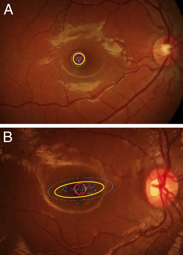
Methods: Participants were 89 children (5-17 years old) with strabismus (n = 31), anisometropia (n = 29), or both conditions (n = 29). Fixation instability was measured using the Nidek MP-1 microperimeter, which calculated horizontal and vertical eye position at 25 Hz as the child attempted steady fixation for 30 seconds. Fixation instability was quantified as the 95% bivariate contour ellipse area (95% BCEA), the best-fit ellipse within which 95% of fixation occurred during the 30-second test. BCEA was normalized by log transformation.
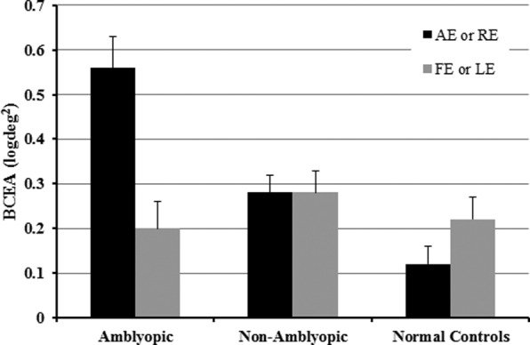
Results: Children with amblyopia had significantly larger BCEAs for amblyopic eyes (mean = 0.56 log deg(2)) than fellow eyes (mean = 0.2 log deg(2), P < 0.01) and right eyes of normal controls (mean = 0.12 log deg(2), P ≤ 0.01). Fixation instability was significantly greater along the horizontal axis of the ellipse for amblyopic (mean = 3.53°) than fellow (mean = 1.98°, P = 0.008), and control (mean = 1.62°, P < 0.001) eyes.
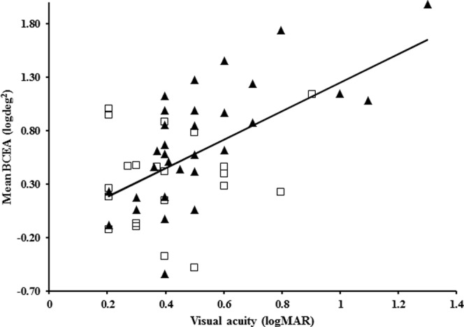
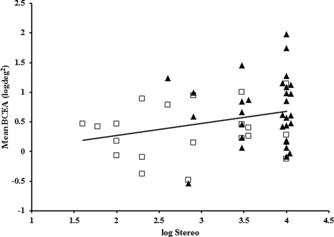
Conclusions: Fixation instability in amblyopic eyes of children with strabismus and/or anisometropia, and the associated poor stereoacuity probably is the consequence of decorrelated binocular experience early in life. Longer duration of decorrelated visual experience is associated with increased fixation instability, poorer stereoacuity, and more severe amblyopia. Treatments that minimize the duration of decorrelated visual experience may improve stereoacuity and decrease fixation instability.
Conflict of interest statement
Disclosure:
Figures
References
-
- Tarita-Nistor L, Gonzalez EG, Markowitz SN, Steinbach MJ. Fixation characteristics of patients with macular degeneration recorded with the mp-1 microperimeter. Retina. 2008; 28: 125–133 - PubMed
-
- Tarita-Nistor L, Gonzalez EG, Mandelcorn MS, Lillakas L, Steinbach MJ. Fixation stability, fixation location, and visual acuity after successful macular hole surgery. Invest Ophthalmol Vis Sci. 2009; 50: 84–89 - PubMed
-
- Gonzalez EG, Tarita-Nistor L, Mandelcorn ED, Mandelcorn M, Steinbach MJ. Fixation control before and after treatment for neovascular age-related macular degeneration. Invest Ophthalmol Vis Sci. 2011; 52: 4208–4213 - PubMed
-
- Regan D, Giaschi DE, Kraft SP, Kothe AC. Method for identifying amblyopes whose reduced line acuity is caused by defective selection and/or control of gaze. Ophthalmic Physiolog Opt. 1992; 12: 425–432 - PubMed
-
- Carpineto P, Ciancaglini M, Nubile M, et al. Fixation patterns evaluation by means of MP-1 microperimeter in microstrabismic children treated for unilateral amblyopia. Eur J Ophthalmol. 2007; 17: 885–890 - PubMed
Publication types
MeSH terms
Grants and funding
LinkOut - more resources
Full Text Sources
Other Literature Sources
Medical
