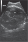
Transcranial Doppler sonographic findings in granulomatous meningoencephalitis in small breed dogs
- PMID: 23372192
- PMCID: PMC3398522
Transcranial Doppler sonographic findings in granulomatous meningoencephalitis in small breed dogs
Abstract
Granulomatous meningoencephalitis (GME) is an acute, progressive, and often fatal inflammatory disease of the central nervous system, affecting mainly small and toy dog breeds. A definitive diagnosis of GME can only be achieved through histopathologic examination of samples collected after death. This retrospective study describes transcranial Doppler ultrasonography (TDS) findings in dogs with confirmed clinical histopathology of GME. Eleven dogs were selected for this study. Sonographic findings in B-mode demonstrated diffuse decreased brain parenchyma echogenicity in 9 dogs, ventriculomegaly in 8 dogs, brain atrophy in 4 dogs, and hyperechoic focal lesions in 6 dogs. Color Doppler imaging revealed more obvious vessels of the arterial circle in 10 dogs. Spectral Doppler examination was performed in 10 dogs to detect the 6 major cerebral arteries of interest. The examination showed normal and high resistive index (RI) values in the outlined arteries. The TDS findings were consistent with pathology found on postmortem examination.
RésuméConstatations échographiques Doppler transcrâniennes pour la méningoencéphalite granulomateuse chez les chiens de petite race. La méningoencéphalite granulomateuse (MEG) est une maladie inflammatoire aiguë, progressive et souvent mortelle du système nerveux central qui touche surtout les chiens de petite race. Un diagnostic définitif de MEG peut seulement être obtenu par un examen histopathologique des échantillons recueillis après la mort. Cette étude rétrospective décrit les constatations d’une échographie Doppler transcrânienne (EDT) chez les chiens avec une histopathologie clinique confirmée de MEG. Onze chiens ont été sélectionnés pour cette étude. Les constatations échographiques en mode-B ont démontré une échogénécité diffuse réduite du parenchyme du cerveau chez 9 chiens, une ventriculomégalie chez 8 chiens, une atrophie du cerveau chez 4 chiens et des lésions focales hyperéchogènes chez 6 chiens. Une imagerie Doppler en couleur a révélé des vaisseaux plus évidents du cercle artériel chez 10 chiens. L’examen spectral Doppler a été réalisé chez 10 chiens pour détecter les 6 artères cérébrales majeures d’intérêt. L’examen a montré des valeurs d’indice de résistivité (IR) normales et hautes dans les artères indiquées. Les constatations de l’EDT étaient conformes à celles de la pathologie trouvées à l’examen postmortem.(Traduit par Isabelle Vallières).
Figures


Similar articles
-
Identification of novel genetic risk loci in Maltese dogs with necrotizing meningoencephalitis and evidence of a shared genetic risk across toy dog breeds.PLoS One. 2014 Nov 13;9(11):e112755. doi: 10.1371/journal.pone.0112755. eCollection 2014. PLoS One. 2014. PMID: 25393235 Free PMC article.
-
Pathological and immunological features of canine necrotising meningoencephalitis and granulomatous meningoencephalitis.Vet J. 2016 Jul;213:72-7. doi: 10.1016/j.tvjl.2016.05.002. Epub 2016 May 5. Vet J. 2016. PMID: 27240919 Review.
-
Comprehensive immunohistochemical studies on canine necrotizing meningoencephalitis (NME), necrotizing leukoencephalitis (NLE), and granulomatous meningoencephalomyelitis (GME).Vet Pathol. 2012 Jul;49(4):682-92. doi: 10.1177/0300985811429311. Epub 2012 Jan 18. Vet Pathol. 2012. PMID: 22262353
-
Clinical presentation and magnetic resonance imaging findings in 11 dogs with eosinophilic meningoencephalitis of unknown aetiology.J Small Anim Pract. 2018 Jul;59(7):422-431. doi: 10.1111/jsap.12837. Epub 2018 Mar 30. J Small Anim Pract. 2018. PMID: 29603737
-
Granulomatous meningoencephalomyelitis in dogs.Compend Contin Educ Vet. 2007 Nov;29(11):678-90. Compend Contin Educ Vet. 2007. PMID: 18210978 Review.
Cited by
-
Transcranial Doppler Ultrasound Examination in Dogs with Suspected Intracranial Hypertension Caused by Neurologic Diseases.J Vet Intern Med. 2018 Jan;32(1):314-323. doi: 10.1111/jvim.14900. Epub 2017 Dec 19. J Vet Intern Med. 2018. PMID: 29265506 Free PMC article.
References
-
- Braund KG. Inflammatory diseases of the central nervous system. In: Braund KG, editor. Clinical Neurology in Small Animals — Localization, Diagnosis and Treatment. Ithaca, New York: International Veterinary Information Service; 2003. [Last accessed June 4, 2012]. pp. 1–49. Available from http://www.ivis.org/special_books/Braund/braund27/ivis.pdf.
-
- Summers BA, Cummings JF, De Lahunta A. Veterinary Neuropathology. 2nd ed. St. Louis, Missouri: Mosby; 1995. Inflammatory diseases of the central nervous system; pp. 110–111.
-
- Muñana KR, Luttgen PJ. Prognostic factors for dogs with granulomatous meningoencephalomyelitis: 42 cases (1982–1996) J Am Vet Med Assoc. 1998;212:1902–1906. - PubMed
MeSH terms
LinkOut - more resources
Full Text Sources
