

Primarily screening and analyzing ESTs differentially expressed in rats' primary liver cancer
- PMID: 23372344
- PMCID: PMC3555298
- DOI: 10.3978/j.issn.1000-9604.2012.12.03
Primarily screening and analyzing ESTs differentially expressed in rats' primary liver cancer
Abstract
Objective: To screen and analyze key express sequence tags (ESTs) which were differentially displayed in every period of SD rats' primary hepatic carcinoma and reveal the molecular mechanism of carcinogenesis.
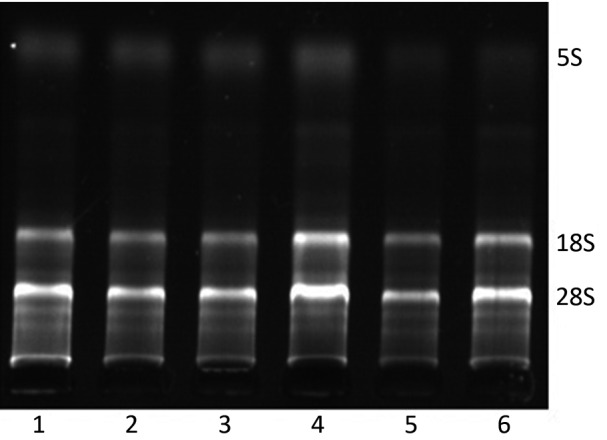
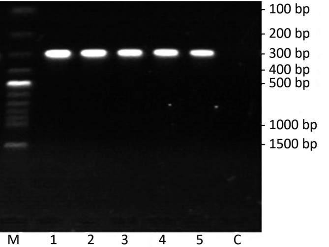
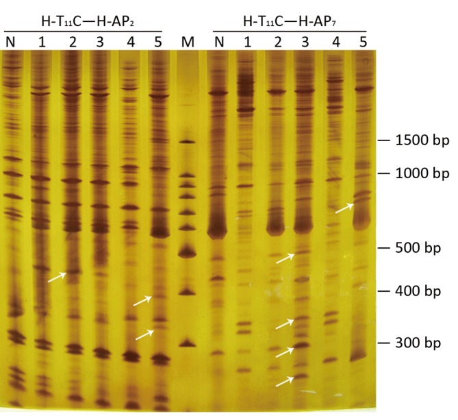
Methods: Using diethylnitrosamine (DENA) as a cancerigenic agent, animal models with different phases of primary hepatic cancer were constructed in SD rats. Rats were respectively sacrificed at d 14, d 28, d 56, d 77, d 105 and d 112 after the rats received DENA by gavage, then the livers were harvested. One part of the livers was classified according to their pathological changes, while the other was reserved for molecular mechanism studies on hepatocarcinogenesis. The differentially expressed genes were isolated from both normal and morbid tissues by mRNA differential display technique (DDRT-PCR). After the fragments were sequenced, bioinformatics were used to analyze the results.
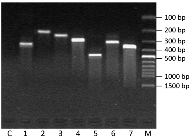
Results: Twelve differentially expressed cDNA fragments were obtained. Nine fragments had the homology with known cDNA clones, especially EST-7 was similar to BN/SsNHsdMCW mitochondrion gene and the identity was 100% which suggested EST-7 may be the part of BN/SsNHsdMCW mitochondrion gene. In contrast, other three fragments (EST-1, EST-3 and EST-5) had extremely low identity to any genes registered in GENBANK databases.
Conclusions: BN/SsNHsdMCW mitochondrion gene was expressed in different periods of hepatocarcinogenesis. Moreover, EST-1, EST-3 and EST-5 were suggested to contribute to the development of rat hepatocarcinogenesis, and thus may be candidates of new targets of oncogenes or cancer suppressor genes.
Keywords: Animal models of primary liver cancer; DDRT-PCR (differential display reverse transcription PCR); ESTs (express sequence tags); mitochondrion gene.
Figures
Similar articles
-
Peanut gene expression profiling in developing seeds at different reproduction stages during Aspergillus parasiticus infection.BMC Dev Biol. 2008 Feb 4;8:12. doi: 10.1186/1471-213X-8-12. BMC Dev Biol. 2008. PMID: 18248674 Free PMC article.
-
Isolation and identification of cDNA fragments and full-length cDNA differentially expressed in human glioblastoma cell line BT-325 versus all-trans retinoic acid induction.Chin Med Sci J. 2000 Dec;15(4):195-200. Chin Med Sci J. 2000. PMID: 12906135
-
Pattern analysis approach reveals restriction enzyme cutting abnormalities and other cDNA library construction artifacts using raw EST data.BMC Biotechnol. 2012 May 3;12:16. doi: 10.1186/1472-6750-12-16. BMC Biotechnol. 2012. PMID: 22554190 Free PMC article.
-
Identification and quantification of differentially expressed transcripts in in vitro-produced bovine preimplantation stage embryos.Mol Reprod Dev. 2003 Oct;66(2):105-14. doi: 10.1002/mrd.10338. Mol Reprod Dev. 2003. PMID: 12950097
-
Meta-analytical biomarker search of EST expression data reveals three differentially expressed candidates.BMC Genomics. 2012;13 Suppl 7(Suppl 7):S12. doi: 10.1186/1471-2164-13-S7-S12. Epub 2012 Dec 13. BMC Genomics. 2012. PMID: 23282184 Free PMC article.
Cited by
-
Mammalian models of chemically induced primary malignancies exploitable for imaging-based preclinical theragnostic research.Quant Imaging Med Surg. 2015 Oct;5(5):708-29. doi: 10.3978/j.issn.2223-4292.2015.06.01. Quant Imaging Med Surg. 2015. PMID: 26682141 Free PMC article. Review.
-
Phytofabrication of Silver nanoparticles: Novel Drug to overcome hepatocellular ailments.Toxicol Rep. 2018 Mar 1;5:333-342. doi: 10.1016/j.toxrep.2018.02.013. eCollection 2018. Toxicol Rep. 2018. PMID: 29854602 Free PMC article.
References
-
- Chuang SC, La Vecchia C, Boffetta P. Liver cancer: descriptive epidemiology and risk factors other than HBV and HCV infection. Cancer Lett 2009;286:9-14 - PubMed
-
- Wang AG, Yoon SY, Oh JH, et al. Identification of intrahepatic cholangiocarcinoma related genes by comparison with normal liver tissues using expressed sequence tags. Biochem Biophys Res Commun 2006;345:1022-32 - PubMed
-
- Shirabe K, Shimada M, Harimoto N, et al. Intrahepatic cholangiocarcinoma: its mode of spreading and therapeutic modalities. Surgery 2002;131:S159-64 - PubMed
-
- Zhang HX, Liu DD, Li L, et al. Screening and identification differentially displayed genes in fore stomach carcinoma of mice. Chin J Cancer Res 2009;21:37-43
-
- Histologic typing of liver tumors of the rat. Institute of Laboratory Animal Resources, National Research Council, National Academy of Sciences, Washington, D.C. J Natl Cancer Inst 1980;64:177-206 - PubMed
LinkOut - more resources
Full Text Sources
Research Materials