

Clinicopathological analysis of metaplastic meningioma: report of 15 cases in Huashan Hospital
- PMID: 23372349
- PMCID: PMC3555304
- DOI: 10.3978/j.issn.1000-9604.2013.01.10
Clinicopathological analysis of metaplastic meningioma: report of 15 cases in Huashan Hospital
Abstract
Objective: Metaplastic meningioma is a rare subtype of benign meningiomas, classified as WHO grade I with well prognosis. Here we presented our experiences on 15 cases of metaplastic meningioma, to investigate the clinicopathological features, therapies and prognosis of these cases.
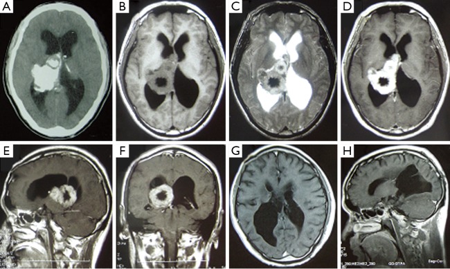
Methods: 15 patients underwent surgical treatment for intracranial metaplastic meningioma between 2001 and 2010 at Neurosurgery Department of Huashan Hospital, Shanghai, China. The clinical data, radiological manifestation, treatment strategy, pathological findings and prognosis of all patients were analyzed retrospectively.
Results: Among the 15 cases (10 males and 5 females), the age ranged from 22 to 74 years old (the mean age was 50.67-year old). The clinical manifestations include headache, dizziness, seizure attack, vision decrease, and weakness of bilateral lower limbs. All the patients received surgical treatment, combined with radiotherapy in some cases. In the follow-up period, recurrence occurred in 2 cases, of which 1 patient died of other system complications.
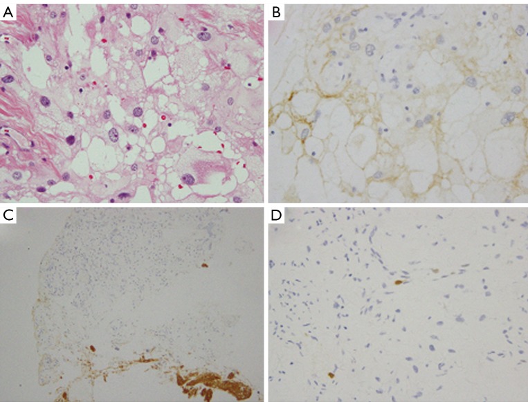
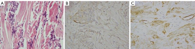
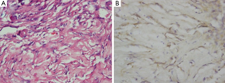
Conclusions: Metaplastic meningiomas are characterized by focal or widespread mesenchymal differentiation with formation of bone, cartilage, fat, and xanthomatous tissue elements. Surgical removal is the optimal therapy, and the overall prognosis is well. But recurrence may occur in some cases, thus radiotherapy is necessary for such kind of patients.
Keywords: Metaplastic meningioma; follow-up; mesenchymal differentiation.
Figures
References
-
- Riemenschneider MJ, Perry A, Reifenberger G. Histological classification and molecular genetics of meningiomas. Lancet Neurol 2006;5:1045-54 - PubMed
-
- Kleihues P, Cavenee WK. eds. World Health Organization Classification of Tumours, Pathology and Genetics of Tumours of the Nervous System. Lyon: IARC, 2000.
-
- Perry A, Louis DN, Scheithauer BW, et al. In: Louis DN, Ohgaki H, Wiestler OD, et al. eds. The WHO Classification of Tumors of the Nervous System. Lyon: IARC, 2007:163-72.
-
- Mawrin C, Perry A.Pathological classification and molecular genetics of meningiomas. J Neurooncol 2010;99:379-91 - PubMed
-
- Roncaroli F, Scheithauer BW, Laeng RH, et al. Lipomatous meningioma: a clinicopathologic study of 18 cases with special reference to the issue of metaplasia. Am J Surg Pathol 2001;25:769-75 - PubMed
LinkOut - more resources
Full Text Sources