

Spectral-domain optical coherence tomography of combined hamartoma of the retina and retinal pigment epithelium in neurofibromatosis
- PMID: 23372386
- PMCID: PMC3550318
- DOI: 10.3341/kjo.2013.27.1.68
Spectral-domain optical coherence tomography of combined hamartoma of the retina and retinal pigment epithelium in neurofibromatosis
Abstract
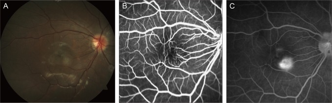
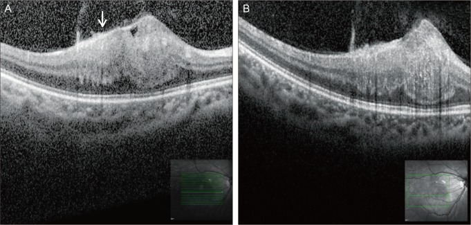
A 5-year-old girl was diagnosed with neurofibromatosis type 2 (NF-2) due to multiple neurofibromas, cafe-au-lait spots, and schwannomas of the brain. During ophthalmologic evaluation, a posterior subcapsular cataract and a gray-green colored subretinal lesion were found in right eye. Fluorescein angiography (FA) revealed a combined hamartoma of the retina and retinal pigment epithelium (CHRRPE). At age 9, she underwent cataract surgery. At this time FA and spectral-domain optical coherence tomography (SD-OCT) were taken. The SD-OCT showed an elevated hyperreflective mass in the retina with prominent attenuation of the inner and outer retina, but minimal attenuation in the photoreceptor layers. The underlying retina appeared to be disorganized and thick (791 µm). This is the first case report of SD-OCT imaging of a CHRRPE associated with NF-2 in a pediatric patient. By using SD-OCT in this patient, we could obtain detailed tumor characteristics, and SD-OCT may be helpful in the diagnosis and management of CHRRPE.
Keywords: Combined hamartoma of retina and retinal pigment epithelium; Neurofibromatoses; Spectral-domain optical coherence tomography.
Conflict of interest statement
No potential conflict of interest relevant to this article was reported.
Figures
Similar articles
-
Association of bilateral, multiple presumed retinal astrocytic proliferations with combined hamartoma of retina and retinal pigment epithelium in a 9-year-old male child with neurofibromatosis type 2.Indian J Ophthalmol. 2016 Nov;64(11):850-852. doi: 10.4103/0301-4738.195609. Indian J Ophthalmol. 2016. PMID: 27958212 Free PMC article.
-
CONCENTRIC MACULAR RINGS SIGN IN COMBINED HAMARTOMA OF RETINA AND RETINAL PIGMENT EPITHELIUM.Retin Cases Brief Rep. 2025 May 1;19(3):333-337. doi: 10.1097/ICB.0000000000001563. Retin Cases Brief Rep. 2025. PMID: 38471086
-
OPTICAL COHERENCE TOMOGRAPHY AND OPTICAL COHERENCE TOMOGRAPHY ANGIOGRAPHY EVALUATION OF COMBINED HAMARTOMA OF THE RETINA AND RETINAL PIGMENT EPITHELIUM.Retina. 2019 May;39(5):1009-1015. doi: 10.1097/IAE.0000000000002053. Retina. 2019. PMID: 29370036
-
Review of spectral domain-enhanced depth imaging optical coherence tomography of tumors of the retina and retinal pigment epithelium in children and adults.Indian J Ophthalmol. 2015 Feb;63(2):128-32. doi: 10.4103/0301-4738.154384. Indian J Ophthalmol. 2015. PMID: 25827543 Free PMC article. Review.
-
Long-term follow-up of adult patient with neurofibromatosis type 1 with retinal astrocytic hamartoma using spectral-domain optical coherence tomography: a review of the literature and a report of a case.Ophthalmic Genet. 2021 Apr;42(2):209-215. doi: 10.1080/13816810.2020.1849315. Epub 2020 Nov 17. Ophthalmic Genet. 2021. PMID: 33203322 Review.
Cited by
-
An Update on the Ophthalmologic Features in the Phakomatoses.J Ophthalmol. 2016;2016:3043026. doi: 10.1155/2016/3043026. Epub 2016 Jul 17. J Ophthalmol. 2016. PMID: 27493794 Free PMC article. Review.
-
Multimodal imaging of combined hamartoma of the retina and retinal pigment epithelium associated with an acquired vitelliform lesion.Int J Retina Vitreous. 2015 Dec 7;1:23. doi: 10.1186/s40942-015-0023-6. eCollection 2015. Int J Retina Vitreous. 2015. PMID: 27847616 Free PMC article.
-
Retinal Imaging of Infants on Spectral Domain Optical Coherence Tomography.Biomed Res Int. 2015;2015:782420. doi: 10.1155/2015/782420. Epub 2015 Jul 6. Biomed Res Int. 2015. PMID: 26221606 Free PMC article. Review.
-
Neurofibromatosis type 2 misdiagnosed as amblyopia-a case report and literature review.Front Med (Lausanne). 2025 Aug 7;12:1556494. doi: 10.3389/fmed.2025.1556494. eCollection 2025. Front Med (Lausanne). 2025. PMID: 40852360 Free PMC article.
References
-
- De Laey JJ, Hanssens M. Vascular tumors and malformations of the ocular fundus. Dordrecht: Kluwer Academic Publishers; 1990. pp. 101–120. - PubMed
-
- Evans DG, Huson SM, Donnai D, et al. A clinical study of type 2 neurofibromatosis. Q J Med. 1992;84:603–618. - PubMed
-
- Pearson-Webb MA, Kaiser-Kupfer MI, Eldridge R. Eye findings in bilateral acoustic (central) neurofibromatosis: association with presenile lens opacities and cataracts but absence of Lisch nodules. N Engl J Med. 1986;315:1553–1554. - PubMed
Publication types
MeSH terms
LinkOut - more resources
Full Text Sources
Other Literature Sources
Medical
Research Materials
Miscellaneous
