Endometriosis, a disease of the macrophage
- PMID: 23372570
- PMCID: PMC3556586
- DOI: 10.3389/fimmu.2013.00009
Endometriosis, a disease of the macrophage
Abstract
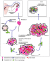
Endometriosis, a common cause of pelvic pain and female infertility, depends on the growth of vascularized endometrial tissue at ectopic sites. Endometrial fragments reach the peritoneal cavity during the fertile years: local cues decide whether they yield endometriotic lesions. Macrophages are recruited at sites of hypoxia and tissue stress, where they clear cell debris and heme-iron and generate pro-life and pro-angiogenesis signals. Macrophages are abundant in endometriotic lesions, where are recruited and undergo alternative activation. In rodents macrophages are required for lesions to establish and to grow; bone marrow-derived Tie-2 expressing macrophages specifically contribute to lesions neovasculature, possibly because they concur to the recruitment of circulating endothelial progenitors, and sustain their survival and the integrity of the vessel wall. Macrophages sense cues (hypoxia, cell death, iron overload) in the lesions and react delivering signals to restore the local homeostasis: their action represents a necessary, non-redundant step in the natural history of the disease. Endometriosis may be due to a misperception of macrophages about ectopic endometrial tissue. They perceive it as a wound, they activate programs leading to ectopic cell survival and tissue vascularization. Clearing this misperception is a critical area for the development of novel medical treatments of endometriosis, an urgent and unmet medical need.
Keywords: Tie-2 expressing macrophages; alternatively activated macrophages; angiogenesis; endometriosis; hypoxia; iron; phagocytosis.
Figures


Similar articles
-
Macrophages are alternatively activated in patients with endometriosis and required for growth and vascularization of lesions in a mouse model of disease.Am J Pathol. 2009 Aug;175(2):547-56. doi: 10.2353/ajpath.2009.081011. Epub 2009 Jul 2. Am J Pathol. 2009. PMID: 19574425 Free PMC article.
-
Differences in growth and vascularization of ectopic menstrual and non-menstrual endometrial tissue in mouse models of endometriosis.Hum Reprod. 2021 Jul 19;36(8):2202-2214. doi: 10.1093/humrep/deab139. Hum Reprod. 2021. PMID: 34109385
-
Basic mechanisms of vascularization in endometriosis and their clinical implications.Hum Reprod Update. 2018 Mar 1;24(2):207-224. doi: 10.1093/humupd/dmy001. Hum Reprod Update. 2018. PMID: 29377994
-
Role of iron overload-induced macrophage apoptosis in the pathogenesis of peritoneal endometriosis.Reproduction. 2014 Jun;147(6):R199-207. doi: 10.1530/REP-13-0552. Epub 2014 Mar 5. Reproduction. 2014. PMID: 24599836 Review.
-
Endometriosis-Associated Macrophages: Origin, Phenotype, and Function.Front Endocrinol (Lausanne). 2020 Jan 23;11:7. doi: 10.3389/fendo.2020.00007. eCollection 2020. Front Endocrinol (Lausanne). 2020. PMID: 32038499 Free PMC article. Review.
Cited by
-
Niclosamide targets the dynamic progression of macrophages for the resolution of endometriosis in a mouse model.Commun Biol. 2022 Nov 11;5(1):1225. doi: 10.1038/s42003-022-04211-0. Commun Biol. 2022. PMID: 36369244 Free PMC article.
-
Identifying Common Pathogenic Features in Deep Endometriotic Nodules and Uterine Adenomyosis.J Clin Med. 2021 Oct 4;10(19):4585. doi: 10.3390/jcm10194585. J Clin Med. 2021. PMID: 34640603 Free PMC article.
-
Transcriptome meta-analysis reveals differences of immune profile between eutopic endometrium from stage I-II and III-IV endometriosis independently of hormonal milieu.Sci Rep. 2020 Jan 15;10(1):313. doi: 10.1038/s41598-019-57207-y. Sci Rep. 2020. PMID: 31941945 Free PMC article.
-
Identification of Shared Molecular Signatures Indicate the Susceptibility of Endometriosis to Multiple Sclerosis.Front Genet. 2018 Feb 16;9:42. doi: 10.3389/fgene.2018.00042. eCollection 2018. Front Genet. 2018. PMID: 29503661 Free PMC article.
-
An in vitro investigation of telocytes-educated macrophages: morphology, heterocellular junctions, apoptosis and invasion analysis.J Transl Med. 2018 Apr 3;16(1):85. doi: 10.1186/s12967-018-1457-z. J Transl Med. 2018. PMID: 29615057 Free PMC article.
References
-
- Allavena P., Chieppa M., Monti P., Piemonti L. (2004). From pattern recognition receptor to regulator of homeostasis: the double-faced macrophage mannose receptor. Crit. Rev. Immunol 24 179–192 - PubMed
LinkOut - more resources
Full Text Sources
Other Literature Sources
Medical
Miscellaneous

