. 2013;8(1):e54715.
doi: 10.1371/journal.pone.0054715. Epub 2013 Jan 25.

Structural and functional analysis of the DEAF-1 and BS69 MYND domains

Affiliations

Structural and functional analysis of the DEAF-1 and BS69 MYND domains

Fatiha Kateb et al. PLoS One. 2013.

Abstract

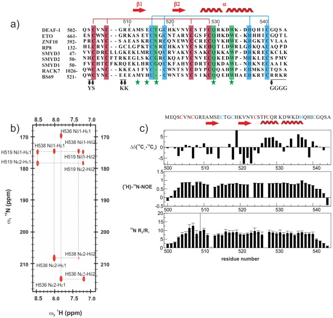
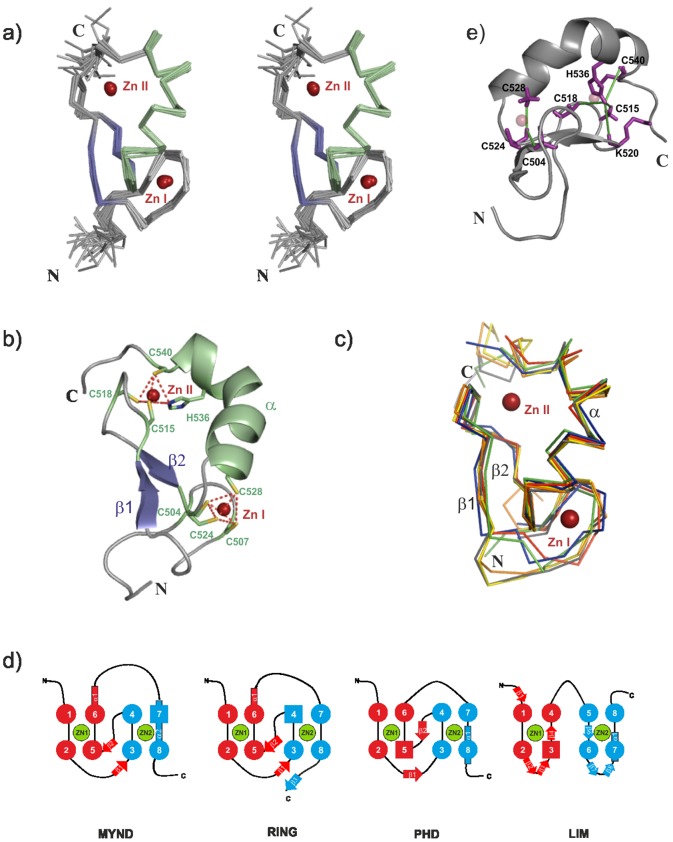
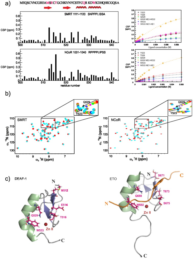
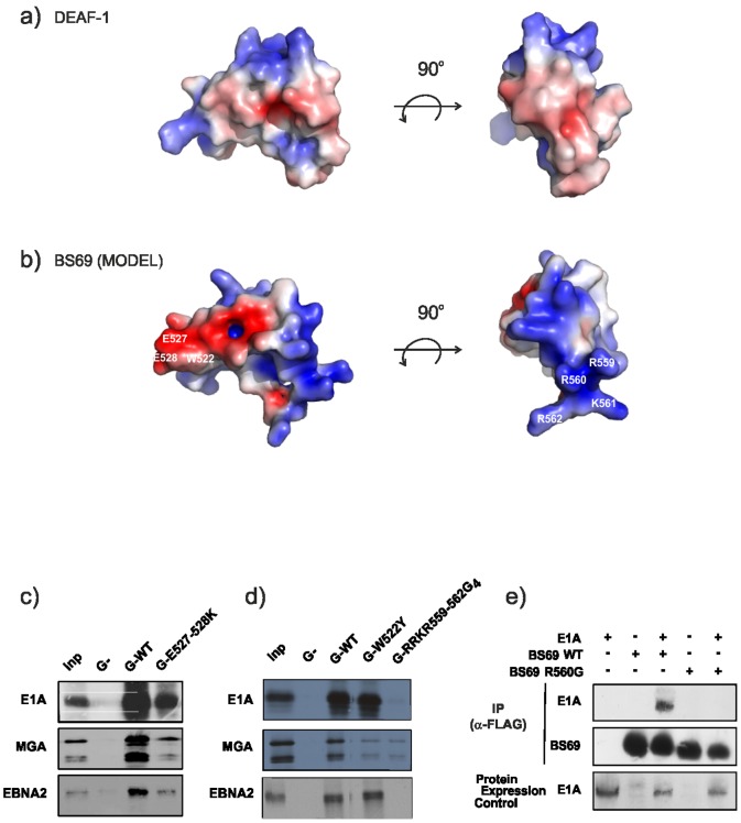
DEAF-1 is an important transcriptional regulator that is required for embryonic development and is linked to clinical depression and suicidal behavior in humans. It comprises various structural domains, including a SAND domain that mediates DNA binding and a MYND domain, a cysteine-rich module organized in a Cys(4)-Cys(2)-His-Cys (C4-C2HC) tandem zinc binding motif. DEAF-1 transcription regulation activity is mediated through interactions with cofactors such as NCoR and SMRT. Despite the important biological role of the DEAF-1 protein, little is known regarding the structure and binding properties of its MYND domain.Here, we report the solution structure, dynamics and ligand binding of the human DEAF-1 MYND domain encompassing residues 501-544 determined by NMR spectroscopy. The structure adopts a ββα fold that exhibits tandem zinc-binding sites with a cross-brace topology, similar to the MYND domains in AML1/ETO and other proteins. We show that the DEAF-1 MYND domain binds to peptides derived from SMRT and NCoR corepressors. The binding surface mapped by NMR titrations is similar to the one previously reported for AML1/ETO. The ligand binding and molecular functions of the related BS69 MYND domain were studied based on a homology model and mutational analysis. Interestingly, the interaction between BS69 and its binding partners (viral and cellular proteins) seems to require distinct charged residues flanking the predicted MYND domain fold, suggesting a different binding mode. Our findings demonstrate that the MYND domain is a conserved zinc binding fold that plays important roles in transcriptional regulation by mediating distinct molecular interactions with viral and cellular proteins.


Conflict of interest statement

Competing Interests: M.J. Bottomley is currently an employee of Novartis Vaccines & Diagnostics. This does not alter the authors' adherence to all the PLOS ONE policies on sharing data and materials.

Figures

Figure 1
Figure 1. Primary sequence and NMR analysis of the DEAF-1 MYND domain.
(a) Sequence alignment of different MYND domains. Residues coordinating the first and second zinc ions are highlighted with red and blue background, respectively. Residues involved in binding to corepressor peptides are indicated with a green star at the bottom, and those interacting through their side chains are highlighted in green. The positions of mutations performed on BS69 are indicated at the bottom. (b) Long range 1H, 15N HSQC spectrum correlating Hε1 and Hδ2 to Nδ1 and Nε2 through 2 J HN and 3 J HN couplings (Pelton et al 1993). The spectrum reveals a different protonation pattern for each histidine sidechain corresponding to the three possible tautomeric states. (c) 13C secondary chemical shifts (top), {1H}-15N heteronuclear NOE (middle), and 15N R2/R1 relaxation rates ratio (bottom) are plotted versus DEAF-1 MYND residue numbers. The secondary structure elements and the amino acid sequence of the protein are indicated at the top of the figure. Residues coordinating the first and second zinc are colored red and blue respectively.
Figure 2
Figure 2. Three-dimensional structure of the DEAF-1 MYND domain.
(a) Stereo view of the ensemble of the twenty lowest energy structures of the DEAF-1 MYND domain. α helices and β strands are colored in green and purple respectively, whereas zinc atoms are depicted as red spheres. (b) Ribbon representation of the DEAF-1 MYND domain. Side-chains of residues coordinating the zinc atoms are shown as sticks. The zinc coordination geometry is indicated by red dotted lines. (c) Superposition of DEAF-1 (green), ETO (red), ZNF10 (Blue), SMYD1 (yellow), SMYD2 (orange) and SMYD3 (gray) MYND structures shown in ribbon representation. The two zinc ions are depicted as red spheres. (d) Schematic representation of the zinc-binding pattern and secondary structure elements in MYND, RING, PHD and LIM domains. (e) Cartoon representation of DEAF1-MYND domain. Side chains of residues for which medium and long-range NOEs are observed that unambiguously define the cross-brace zinc binding topology are shown in magenta. Green lines indicate NOEs between C524 HN/C504 Hβ*, C524 HN/C528 Hβ* for the first binding site; and H536 Hε1/C540 HN, H536 Hε1/C518 Hβ1, H536 Hε1/K520 HN, and H536 Hε1/C515 Hβ2 for the second binding site.
Figure 3
Figure 3. Binding of DEAF-1 MYND to SMRT and NCoR peptides.
(a) Chemical Shift Perturbations (CSP, see methods) observed for the interaction between DEAF-1 MYND domain and SMRT (top) and NCoR (bottom) corepressor peptides. The sequence of SMRT and NCoR peptides used for the titrations are indicated in each graph. Secondary structure elements and amino acid sequence of the DEAF-1 MYND domain are shown at the top of the panel. Residues expected to interact with the corepressors are colored magenta. CSPs of residues that are most strongly affected are shown (cross symbols) as a function of ligand concentration for both titration with SMRT (top right) and NCoR (bottom right). The observed CSPs were fitted to a binding isotherm yielding dissociation constanst of 5.30±0.54 mM and 3.08±0.12 mM for the SMRT and NCoR peptides, respectively. The binding curves are shown as dashed lines. (b) Superposition of 1H, 15N HSQC spectra of a 1 mM sample of the free DEAF-1 MYND domain (red) and upon addition of unlabeled corepressor peptides (cyan) up to a final concentration of 8 mM and 5 mM of SMRT and NCoR peptide (1∶8 and 1∶5 molar ratio), respectively. The intermediate steps of each titration are zoomed for a sub-region of the corresponding spectrum. In either case binding takes place on the fast exchange regime with respect to the chemical shift time scale. (c) Ribbon representation of DEAF-1 (left) and ETO (right) MYND domains. Residues experiencing the largest chemical shift perturbation upon addition of the corepressor peptides are shown as magenta sticks. In the ETO-SMRT complex structure the corresponding residues are shown as sticks as well, and the SMRT ligand peptide is colored orange and shown in cartoon representation.
Figure 4
Figure 4. Binding studies and mutational analysis of BS69 MYND domain.
(a) Two views of an electrostatic surface representation of the DEAF-1 MYND domain. Positive (blue) and negative (red) electrostatic surface potential is shown at ±3 kB T/e and was determined using the program APBS in Pymol (www.pymol.org). (b) Corresponding surface views for the BS69 MYND structure obtained from homology modelling. Positive (blue) and negative (red) electrostatic surface potential is displayed at ±3 kB T/e. The location of the residues that were mutated for binding studies are labelled on the surface representation of the BS69 homology model. (c),(d) Analysis of binding of E1A, EBNA2 and MGA to wild-type or mutant BS69 proteins expressed as a GST-fusion protein or to the GST (G−) moiety as control; Inp: Input 10%; G-RRKR-559-562G4: mutation of residues 559–562 into four glycines. (e) A single point mutation in BS69 abrogates the binding to E1A. QT6 fibroblasts were transfected with 12SE1A and/or FLAG-tagged BS69 expression vectors as indicated. Cellular complexes were immunoprecipitated with an M2 anti-FLAG antibody. Co-immunoprecipitated and ectopic expressions of E1A were revealed by immunoblotting using an M73-E1A antibody.

Similar articles

Cited by

  • Mutations affecting the SAND domain of DEAF1 cause intellectual disability with severe speech impairment and behavioral problems.
    Vulto-van Silfhout AT, Rajamanickam S, Jensik PJ, Vergult S, de Rocker N, Newhall KJ, Raghavan R, Reardon SN, Jarrett K, McIntyre T, Bulinski J, Ownby SL, Huggenvik JI, McKnight GS, Rose GM, Cai X, Willaert A, Zweier C, Endele S, de Ligt J, van Bon BW, Lugtenberg D, de Vries PF, Veltman JA, van Bokhoven H, Brunner HG, Rauch A, de Brouwer AP, Carvill GL, Hoischen A, Mefford HC, Eichler EE, Vissers LE, Menten B, Collard MW, de Vries BB. Vulto-van Silfhout AT, et al. Am J Hum Genet. 2014 May 1;94(5):649-61. doi: 10.1016/j.ajhg.2014.03.013. Epub 2014 Apr 10. Am J Hum Genet. 2014. PMID: 24726472 Free PMC article.
  • Zn-regulated GTPase metalloprotein activator 1 modulates vertebrate zinc homeostasis.
    Weiss A, Murdoch CC, Edmonds KA, Jordan MR, Monteith AJ, Perera YR, Rodríguez Nassif AM, Petoletti AM, Beavers WN, Munneke MJ, Drury SL, Krystofiak ES, Thalluri K, Wu H, Kruse ARS, DiMarchi RD, Caprioli RM, Spraggins JM, Chazin WJ, Giedroc DP, Skaar EP. Weiss A, et al. Cell. 2022 Jun 9;185(12):2148-2163.e27. doi: 10.1016/j.cell.2022.04.011. Epub 2022 May 17. Cell. 2022. PMID: 35584702 Free PMC article.
  • Refining analyses of copy number variation identifies specific genes associated with developmental delay.
    Coe BP, Witherspoon K, Rosenfeld JA, van Bon BW, Vulto-van Silfhout AT, Bosco P, Friend KL, Baker C, Buono S, Vissers LE, Schuurs-Hoeijmakers JH, Hoischen A, Pfundt R, Krumm N, Carvill GL, Li D, Amaral D, Brown N, Lockhart PJ, Scheffer IE, Alberti A, Shaw M, Pettinato R, Tervo R, de Leeuw N, Reijnders MR, Torchia BS, Peeters H, O'Roak BJ, Fichera M, Hehir-Kwa JY, Shendure J, Mefford HC, Haan E, Gécz J, de Vries BB, Romano C, Eichler EE. Coe BP, et al. Nat Genet. 2014 Oct;46(10):1063-71. doi: 10.1038/ng.3092. Epub 2014 Sep 14. Nat Genet. 2014. PMID: 25217958 Free PMC article.
  • BS69/ZMYND11 C-Terminal Domains Bind and Inhibit EBNA2.
    Harter MR, Liu CD, Shen CL, Gonzalez-Hurtado E, Zhang ZM, Xu M, Martinez E, Peng CW, Song J. Harter MR, et al. PLoS Pathog. 2016 Feb 4;12(2):e1005414. doi: 10.1371/journal.ppat.1005414. eCollection 2016 Feb. PLoS Pathog. 2016. PMID: 26845565 Free PMC article.
  • DEAF1 binds unmethylated and variably spaced CpG dinucleotide motifs.
    Jensik PJ, Vargas JD, Reardon SN, Rajamanickam S, Huggenvik JI, Collard MW. Jensik PJ, et al. PLoS One. 2014 Dec 22;9(12):e115908. doi: 10.1371/journal.pone.0115908. eCollection 2014. PLoS One. 2014. PMID: 25531106 Free PMC article.

References

    1. Gross CT, McGinnis W (1996) DEAF-1, a novel protein that binds an essential region in a Deformed response element. EMBO J 15: 1961–1970. - PMC - PubMed
    1. Michelson RJ, Collard MW, Ziemba AJ, Persinger J, Bartholomew B, et al. (1999) Nuclear DEAF-1-related (NUDR) Protein Contains a Novel DNA Binding Domain and Represses Transcription of the Heterogeneous Nuclear Ribonucleoprotein A2/B1 Promoter. JBC 274: 30510–30519. - PubMed
    1. Veraksa A, Kennison J, McGinnis W (2002) DEAF-1 Function Is Essential for the Early Embryonic Development of Drosophila. genesis 33: 67–76. - PubMed
    1. Lemonde S, Turecki G, Bqkish D, Du L, Hrdina PD, et al. (2003) Impaired Repression at a 5-Hydroxytryptamine 1A Receptor Gene Polymorphism Associated with Major Depression and Suicide. J Neurosci 23: 8788–8799. - PMC - PubMed
    1. Barker HE, Smyth GK, Wettenhall J, Ward TA, Bath ML, et al. (2008) Deaf-1 regulates epithelial cell proliferation and side-branching in the mammary gland. BMC Dev Biol 8. - PMC - PubMed

Publication types

MeSH terms

Associated data

LinkOut - more resources