. 2013;8(1):e54799.
doi: 10.1371/journal.pone.0054799. Epub 2013 Jan 25.

Atad3 function is essential for early post-implantation development in the mouse

Affiliations

Atad3 function is essential for early post-implantation development in the mouse

Tobias Goller et al. PLoS One. 2013.

Abstract

The mitochondrial AAA+-ATPase ATAD3 is implicated in the regulation of mitochondrial and ER dynamics and was shown to be necessary for larval development in Caenorhabditis elegans. In order to elucidate the relevance of ATAD3 for mammalian development, the phenotype of an Atad3 deficient mouse line was analyzed. Atad3 deficient embryos die around embryonic day E7.5 due to growth retardation and a defective development of the trophoblast lineage immediately after implantation into the uterus. This indicates an essential function of Atad3 for the progression of the first steps of post-implantation development at a time point when mitochondrial biogenesis and ATP production by oxidative phosphorylation are required. Therefore, murine Atad3 plays an important role in the biogenesis of mitochondria in trophoblast stem cells and in differentiating trophoblasts. At the biochemical level, we report here that ATAD3 is present in five native mitochondrial protein complexes of different sizes, indicating complex roles of the protein in mitochondrial architecture and function.


Conflict of interest statement

Competing Interests: The authors have declared that no competing interests exist.

Figures

Figure 1
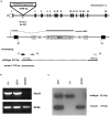
Figure 1. Gene trap mutagenesis of the murine Atad3 locus.
A Schemes of the murine Atad3 genomic locus on chromosome 4 showing the integration site of the gene trap vector rFlipROSAbetageo(Cre)0 into intron 1 at position 402 as an overview (upper scheme) and in detail (middle scheme), as well as the positions of the genotyping primers and resulting amplificates (lower scheme). Boxes in the upper scheme represent all 16 exons, the untranslated regions (UTRs) are without filling. In the middle scheme it is depicted, that the gene trap vector insertion leads to a fusion of 5? exon 1 (E1) to the splice acceptor side (SA) of rFlipROSAbetageo(Cre)0. The mutant mRNA contains Atad3 exon 1 and the vector βgeo cassette and is terminated by an SV40 polyadenylation signal (pA). Retroviral long terminal repeat (LTR) sequences are necessary for efficient vector integration. Recognition sites (rec) for Flp and Cre recombination enzymes allow first the reconstitution of the wildtype mRNA and secondly a subsequent conditional mutagenesis. In the lower scheme, arrows indicate the positions of the PCR primers EF (endogenous intron 1 forward primer), ER (endogenous intron 1 reverse primer) and VR (gene trap vector reverse primer) used for genotyping. Constitution of wildtype (813 bp) and mutant (273 bp) PCR fragments are shown below. B RT-PCR analysis of E6.5 embryonic samples shows that the complete 3? region (exons 11 to 16) of the Atad3 cDNA is missing in Atad3GT/GT embryos, instead in wildtype and Atad3G/+ tissues a 805 bp fragment represents the Atad3 cDNA. Amplification of ribosomal protein S6 (RPS6) cDNA serves as quantity control. C For genotyping of the Atad3 locus, wildtype and mutant genomic PCR amplificates are separated in a 1.5% agarose gel and identify samples from wildtype (+/+), heterozygous (GT/+) and homozygous mutant (GT/GT) individuals.
Figure 2
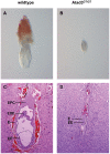
Figure 2. Atad3GT/GT embryos are defective in early post-implantation development.
A, B Morphology of a wildtype (A) and an Atad3GT/GT (B) embryo at E6.5. Atad3GT/GT embryos show a total growth reduction, especially the proximo-distal axis is not extended. C, D Anatomical analysis of a wildtype (C) and an Atad3GT/GT (D) embryo at E7.5 by hematoxilin/eosin staining of 7 µm paraffin sections (same magnifications). In Atad3GT/GT embryos EPC and EXE are strongly reduced. Additionally, EE and E appear less differentiated. Abbreviations: E endoderm, EE embryonic ectoderm, EPC ectoplacental cone, EXE extra-embryonic ectoderm.
Figure 3
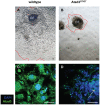
Figure 3. Atad3GT/GT embryos are defective in trophoblast development.
A, B Morphology of a wildtype (A) and an Atad3GT/GT (B) embryo outgrowth, isolated at E6.5, and after 3 days of in?vitro cultivation. Red lines mark the borders of the outgrowths. Compared to the wildtype tissues, the size of the epiblast remains clearly diminished in Atad3GT/GT embryos. EE epiblast.C, D Atad3 protein expression and mitochondrial morphology in the middle region of a wildtype trophoblast outgrowth (C) and a complete Atad3GT/GT (D) embryo outgrowth are shown by immuno-staining with an Atad3 specific antibody (green). Nuclei are visualized by DAPI staining (blue). In Atad3GT/GT outgrowths, Atad3 protein is only weakly expressed in the epiblasts and is almost undetectable in the trophoblast. The white arrow in D indicates the epiblast.
Figure 4
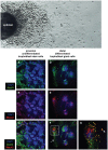
Figure 4. Alterations in mitochondrial morphology during trophoblast differentiation.
The overview picture shows the different areas of a typical E6.5 embryo outgrowth (outgrowth direction from left to right): the epiblast mountain (left), followed by the proximal zone of densely arranged undifferentiated trophoblast stem cells and finally the distal zone (right) of differentiated trophoblast cells. A–G The alteration of mitochondrial morphology during proximo-distal trophoblast differentiation in wildtype outgrowths is shown by co-immunostaining of the mitochondrial marker Atad3 (green) and the trophoblast differentiation marker Mash2 (red). Nuclei are visualized by DAPI staining (blue). In cells of the proximal trophoblast (A – C), the mitochondria are small and diffusely distributed (A), here Mash2 is localized to the cytoplasm (B). Instead in the distal region of the outgrowths (D – G), the cells contain enlarged, swollen mitochondria (D, F and arrowhead in G), and additionally small mitochondria arranged in arrays from the center to the periphery of the cell (D, arrow in F). In distal cells, Mash2 is localized to the nucleus and is also detected in the matrix of the swollen mitochondria (E, F and G). G is a magnified detail of F.
Figure 5
Figure 5. Atad3GT/GT embryos are defective in trophoblast differentiation.
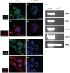
A, B Intrinsic apoptosis in wildtype (A) and Atad3GT/GT (B) embryo outgrowths at E6.5 was analyzed by immuno-staining for cytochrome c (red). Mitochondria in wildtype (A) and Atad3GT/GT (B) cells are marked by Atad3 immuno-staining (green). A punctate cytochrome c expression pattern is seen in wildtype (A’) as well as in Atad3GT/GT cells (B’). C, D Differentiated trophoblast cells in in the most distal region of wildtype (C) and Atad3GT/GT (D) embryo outgrowths at E6.5 were detected by immuno-staining for Mash2 (red). Mitochondria in wildtype (C) and Atad3GT/GT (D) cells are marked by Atad3 immuno-staining (green). Analysis was performed by confocal microscopy. In contrast to wildtype cells (C’), Mash2 is rarely detectable in cells of the most distal region of the Atad3GT/GT trophoblast outgrowth (D’). All nuclei are visualized by DAPI staining (blue). E The expression of trophoblast cell type specific marker genes in E6.5 wildtype and Atad3GT/GT embryos was monitored by RT-PCR. As opposed to strong signals for all analysed markers in the wildtype sample, in the Atad3GT/GT specimen Cdx2 and Bmp4 transcripts are not detected and Mash2 and Hand1 amplificates are observed at very low levels.
Figure 6
Figure 6. ATAD3 contributes to five mitochondrial protein complexes of different sizes.
A The expression of human ATAD3A/Atad3 in different mitochondrial protein complexes in HeLa cells (first lane) and in the murine ES cells (second lane) is shown by Blue native PAGE (gradient of acrylamide concentration: 3–12%) and subsequent immuno-blotting for ATAD3A. In both cell lines the same expression pattern is seen. Both lanes depicted have run on the same gel and are taken from the same ATAD3A immuno-blot. B In HeLa cells, ATAD3A is detected in five different protein complexes (first lane). The largest and most abundant complex (main complex) has a molecular weight of about 800 to 900 kDa. The four smaller sub-complexes I–IV have estimated molecular weights of about 720, 600, 480 and 240 kDa. In HeLa cells, Blue native PAGE (gradient of acrylamide concentration: 3–12%) and subsequent immuno-blotting shows that the mitochondrial fission and fusion proteins MFN1, MFN2 and DRP1 are contained in protein complexes of similar molecular weights as ATAD3A sub-complexes III and IV (second to fourth lane). All four lanes depicted have run on the same gel. Sizes of the protein standard refer to IgM hexamer (1236 kDa), IgM pentamer (1048 kDa), Apoferritin band 1 (720 kDa), Apoferritin band 2 (480 kDa) and B-phycoerythrin (242 kDa).

References

    1. Schaffrik M, Mack B, Matthias C, Rauch J, Gires O (2006) Molecular characterization of the tumor-associated antigen AAA-TOB3. Cell Mol Life Sci 63: 2162–2174. - PMC - PubMed
    1. Gilquin B, Taillebourg E, Cherradi N, Hubstenberger A, Gay O, et al. (2010) The AAA+ ATPase ATAD3A controls mitochondrial dynamics at the interface of the inner and outer membranes. Mol Cell Biol 30: 1984–96. - PMC - PubMed
    1. Neuwald AF, Aravind L, Spouge JL, Koonin EV (1999) AAA+: A class of chaperone-like ATPases associated with the assembly, operation, and disassembly of protein complexes. Genome Res 9: 27–43. - PubMed
    1. Langer T, Käser M, Klanner C, Leonhard K (2001) AAA proteases of mitochondria: quality control of membrane proteins and regulatory functions during mitochondrial biogenesis. Biochem Soc Trans 29: 431–6. - PubMed
    1. Hanson PI, Whiteheart SW (2005) AAA+ proteins: have engine, will work. Nat Rev Mol Cell Biol 6: 519–29. - PubMed

Publication types

MeSH terms

Substances