Molecular analysis of precursor lesions in familial pancreatic cancer
- PMID: 23372777
- PMCID: PMC3553106
- DOI: 10.1371/journal.pone.0054830
Molecular analysis of precursor lesions in familial pancreatic cancer
Abstract
Background: With less than a 5% survival rate pancreatic adenocarcinoma (PDAC) is almost uniformly lethal. In order to make a significant impact on survival of patients with this malignancy, it is necessary to diagnose the disease early, when curative surgery is still possible. Detailed knowledge of the natural history of the disease and molecular events leading to its progression is therefore critical.
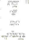
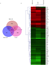
Methods and findings: We have analysed the precursor lesions, PanINs, from prophylactic pancreatectomy specimens of patients from four different kindreds with high risk of familial pancreatic cancer who were treated for histologically proven PanIN-2/3. Thus, the material was procured before pancreatic cancer has developed, rather than from PanINs in a tissue field that already contains cancer. Genome-wide transcriptional profiling using such unique specimens was performed. Bulk frozen sections displaying the most extensive but not microdissected PanIN-2/3 lesions were used in order to obtain the holistic view of both the precursor lesions and their microenvironment. A panel of 76 commonly dysregulated genes that underlie neoplastic progression from normal pancreas to PanINs and PDAC were identified. In addition to shared genes some differences between the PanINs of individual families as well as between the PanINs and PDACs were also seen. This was particularly pronounced in the stromal and immune responses.
Conclusions: Our comprehensive analysis of precursor lesions without the invasive component provides the definitive molecular proof that PanIN lesions beget cancer from a molecular standpoint. We demonstrate the need for accumulation of transcriptomic changes during the progression of PanIN to PDAC, both in the epithelium and in the surrounding stroma. An identified 76-gene signature of PDAC progression presents a rich candidate pool for the development of early diagnostic and/or surveillance markers as well as potential novel preventive/therapeutic targets for both familial and sporadic pancreatic adenocarcinoma.
Conflict of interest statement
Figures


Similar articles
-
Genetic alterations associated with progression from pancreatic intraepithelial neoplasia to invasive pancreatic tumor.Gastroenterology. 2013 Nov;145(5):1098-1109.e1. doi: 10.1053/j.gastro.2013.07.049. Epub 2013 Aug 2. Gastroenterology. 2013. PMID: 23912084 Free PMC article.
-
Molecular genetics of pancreatic intraepithelial neoplasia.J Hepatobiliary Pancreat Surg. 2007;14(3):224-32. doi: 10.1007/s00534-006-1166-5. Epub 2007 May 29. J Hepatobiliary Pancreat Surg. 2007. PMID: 17520196 Free PMC article. Review.
-
The biological features of PanIN initiated from oncogenic Kras mutation in genetically engineered mouse models.Cancer Lett. 2013 Oct 1;339(1):135-43. doi: 10.1016/j.canlet.2013.07.010. Epub 2013 Jul 22. Cancer Lett. 2013. PMID: 23887057
-
Annexin A10 is a candidate marker associated with the progression of pancreatic precursor lesions to adenocarcinoma.PLoS One. 2017 Apr 3;12(4):e0175039. doi: 10.1371/journal.pone.0175039. eCollection 2017. PLoS One. 2017. PMID: 28369074 Free PMC article.
-
Pancreatic intraepithelial neoplasia-can we detect early pancreatic cancer?Histopathology. 2010 Oct;57(4):503-14. doi: 10.1111/j.1365-2559.2010.03610.x. Epub 2010 Sep 28. Histopathology. 2010. PMID: 20875068 Review.
Cited by
-
Applications of gene pair methods in clinical research: advancing precision medicine.Mol Biomed. 2025 Apr 9;6(1):22. doi: 10.1186/s43556-025-00263-w. Mol Biomed. 2025. PMID: 40202606 Free PMC article. Review.
-
Pancreatic preneoplastic lesions plasma signatures and biomarkers based on proteome profiling of mouse models.Br J Cancer. 2015 Dec 1;113(11):1590-8. doi: 10.1038/bjc.2015.370. Epub 2015 Oct 29. Br J Cancer. 2015. PMID: 26512875 Free PMC article.
-
Amino acid transporter SLC6A14 is a novel and effective drug target for pancreatic cancer.Br J Pharmacol. 2016 Dec;173(23):3292-3306. doi: 10.1111/bph.13616. Epub 2016 Oct 18. Br J Pharmacol. 2016. PMID: 27747870 Free PMC article.
-
Identification of Key Genes and Pathways in Pancreatic Cancer Gene Expression Profile by Integrative Analysis.Genes (Basel). 2019 Aug 13;10(8):612. doi: 10.3390/genes10080612. Genes (Basel). 2019. PMID: 31412643 Free PMC article.
-
NAD+ metabolism governs the proinflammatory senescence-associated secretome.Nat Cell Biol. 2019 Mar;21(3):397-407. doi: 10.1038/s41556-019-0287-4. Epub 2019 Feb 18. Nat Cell Biol. 2019. PMID: 30778219 Free PMC article.
References
-
- Jemal A, Siegel R, Ward E, Hao Y, Xu J, et al. (2008) Cancer statistics, 2008. CA Cancer J Clin 58: 71–96. - PubMed
-
- Hruban RH, Adsay NV, Albores-Saavedra J, Compton C, Garrett ES, et al. (2001) Pancreatic intraepithelial neoplasia: a new nomenclature and classification system for pancreatic duct lesions. Am J Surg Pathol 25: 579–586. - PubMed
-
- Real FX, Cibrian-Uhalte E, Martinelli P (2008) Pancreatic cancer development and progression: remodeling the model. Gastroenterology 135: 724–728. - PubMed
-
- Buchholz M, Braun M, Heidenblut A, Kestler HA, Kloppel, G, et al. ( 2005) Transcriptome analysis of microdissected pancreatic intraepithelial neoplastic lesions. Oncogene 24: 6626–6636. - PubMed
Publication types
MeSH terms
Substances
Supplementary concepts
Associated data
- Actions
Grants and funding
LinkOut - more resources
Full Text Sources
Other Literature Sources
Medical
Molecular Biology Databases

