. 2013;8(1):e55055.
doi: 10.1371/journal.pone.0055055. Epub 2013 Jan 25.

The anaphase promoting complex contributes to the degradation of the S. cerevisiae telomerase recruitment subunit Est1p

Affiliations

The anaphase promoting complex contributes to the degradation of the S. cerevisiae telomerase recruitment subunit Est1p

Jenifer L Ferguson et al. PLoS One. 2013.

Abstract

Telomerase is a multi-subunit enzyme that reverse transcribes telomere repeats onto the ends of linear eukaryotic chromosomes and is therefore critical for genome stability. S. cerevisiae telomerase activity is cell-cycle regulated; telomeres are not elongated during G1 phase. Previous work has shown that Est1 protein levels are low during G1 phase, preventing telomerase complex assembly. However, the pathway targeting Est1p for degradation remained uncharacterized. Here, we show that Est1p stability through the cell cycle mirrors that of Clb2p, a known target of the Anaphase Promoting Complex (APC). Indeed, Est1p is stabilized by mutations in both essential and non-essential components of the APC. Mutations of putative Destruction boxes (D-boxes), regions shown to be important for recognition of known APC substrates, stabilize Est1p, suggesting that Est1p is likely to be targeted for degradation directly by the APC. However, we do not detect degradation or ubiquitination of recombinant Est1p by the APC in vitro, suggesting either that the recombinant protein lacks necessary post-translational modification and/or conformation, or that the APC affects Est1p degradation by an indirect mechanism. Together, these studies shed light on the regulation of yeast telomerase assembly and demonstrate a new connection between telomere maintenance and cell cycle regulation pathways.


Conflict of interest statement

Competing Interests: The authors have declared that no competing interests exist.

Figures

Figure 1
Figure 1. Est1p is unstable in G1 phase, but stable in early S and G2/M phases.
(A) Endogenously expressed Est1p-MYC13p levels during cell cycle arrests. Strains YKF800 (untagged; lane 1) and YKF801 (EST1-MYC13; lanes 2–5) were grown asynchronously at 30°C to mid-log phase and then left untreated (asynchronous) or arrested by addition of α-factor, hydroxyurea, or nocodazole, as indicated. When 95% of the population was arrested, as monitored by the bud-index, cells were harvested. Whole-cell extract was prepared and western blotted using anti-MYC, anti-Clb2p, and anti-Actin antibodies, as indicated. (B) Half-life of HA3-Est1p during cell cycle arrests. Strain YKF802 containing plasmid pVL242RtoA (PGAL1-HA3-EST1) was grown asynchronously at 30°C to mid-log phase and arrested with α-factor, hydroxyurea, or nocodazole, as indicated. When 95% of the population was arrested, as monitored by the bud-index, expression of HA3-EST1 was induced with addition of galactose and then subsequently repressed (after 1 hour) with glucose and cycloheximide (time 0). Samples from cells harvested at the indicated times were western blotted with anti-HA, anti-Clb2p and anti-Actin antibodies, as indicated. An induced asynchronous sample of strain YKF806+ pVL242RtoA (clb2Δ; left panel), served as a negative control for Clb2p and positive control for HA3-Est1p detection. An uninduced asynchronous sample of strain YKF802+ pVL242RtoA (Raff; left panel) served as a positive control for Clb2p detection and negative control for HA3-Est1p specificity. A non-specific background band is indicated by formula image. (C) Quantification of data shown in (B), as described in Materials and Methods. The calculated half-lives were averaged from independent biological replicates: αF (α-factor), n = 7; HU (hydroxyurea), n = 4; NOC (nocodazole), n = 4. Error bars are standard deviation from the mean. Both HU and NOC are statistically different from αF by two-tailed t-test (p-values 1.1×10⁻⁵ and 1.1×10⁻⁶, respectively) as denoted by *.
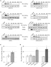
Figure 2
Figure 2. APC function is required for normal Est1p degradation during G1 phase.
(A) HA3-Est1p stability increases when APC function is compromised. Western blots of Est1p stability assays from strain K4438 (cdc16-123) harboring pKF600 (GAL1-HA3-EST1) plus either a complementing vector pRS416-CDC16 (labeled “CDC16”) or an empty vector pRS416 (labeled “cdc16-123”) were conducted as described in Materials and Methods. An uninduced sample (Raff) served as a negative control for HA3-Est1p specificity. (B) HA3-Est1p is stabilized in APC deletion mutants. Western blots of Est1p stability assays from strains YKF802 (Wild Type), YKF803 (apc9Δ), YKF804 (mnd2Δ), YKF805 (swm1Δ), YKF806 (clb2Δ) and YKF807 (clb2Δcdh1Δ) containing pVL242RtoA (PGAL1-HA3-EST1) were conducted as described in Materials and Methods. For YKF805 (swm1Δ), an uninduced asynchronous sample (Raff) served as a positive control for Clb2p detection and negative control for HA3-Est1p specificity, while an uninduced asynchronous sample of strain YKF806 (clb2Δ) served as a negative control for Clb2p detection. (C) Quantification of results shown in (A). Bars represent the average HA3-Est1p half-life from three independent biological replicates. Error bars are standard deviation of the mean (p-value = 0.08 by two-tailed t test). (D) Quantification of results shown in (B). Bars represent the average HA3-Est1p half-life from independent biological replicates: n = 3 for all strains except clb2Δcdh1Δ, where n = 4. Error bars are standard deviation from the mean. By two-tailed paired t-test, there is a significant difference between the control (WT) and swm1Δ (p-value 0.0002) but not between WT and apc9Δ (p-value 0.49) or mnd2Δ (p-value 0.83). There is a significant difference between the control (clb2Δ) and clb2Δcdh1Δ strains (p-value 0.003). Significant differences are denoted by *.
Figure 3
Figure 3. Cell-cycle oscillation of Est1p requires Cdh1p.
(A) Est1 protein levels oscillate through the cell cycle. Strain YKF808 (cdc15-2 EST1-MYC13) was grown asynchronously at 23°C to mid-log phase and shifted to the restrictive temperature (37°C) for 3.5 hrs. When 95% of the cells were arrested, as monitored by bud-index (Figure S2), the culture was returned to the permissive temperature (23°C; time 0). Whole-cell extract was prepared from samples harvested every 20 mins following release and western blotted using anti-MYC, anti-Clb2p, and anti-Actin antibodies, as indicated. YCM191 (cdc15-2) served as the untagged (No MYC) control for Est1-MYC13p and was harvested following the 37°C incubation period. Est1-MYC13p and Clb2p intensity at each time were normalized to input (actin) and starting amount (time 0). Bars represent the average of four independent biological replicates for Est1-MYC13p (light) and Clb2p (dark); error bars are standard deviation of the mean. (B) Deletion of CDH1 perturbs the oscillation of Est1-MYC13p through the cell cycle. Strain YKF809 (cdc15-2 cdh1Δ EST1-MYC13) was treated as in (A), except the bars represent the average of three independent biological replicates.
Figure 4
Figure 4. Est1p degradation in G1 phase requires three destruction boxes (D-boxes).
(A) Schematic of EST1 shown to scale. EST1 contains six putative D-boxes with sequence RxxL (boxes labeled 1–6). Deletion of the C-terminal 300 amino acids (CΔ300) results in a truncated protein that removes putative D-boxes 5 and 6. The N-terminal 52 amino acids are shown, with putative D-boxes 1 and 2 outlined. Upward pointing black triangles represent the position of the indicated N-terminal deletion. (B) D-boxes 1, 2, and 4 contribute to Est1p degradation. YKF802 containing pKF600 (GAL1-HA3-EST1) plasmids expressing either wild-type EST1 (WT) or the D-box (DB) mutated (RxxL to AxxA) est1 alleles indicated were treated as in Figure 1B, except strains were arrested with α-factor. Bars represent the average HA3-Est1p half-life for three independent biological replicates; error bars are the standard deviation of the mean. Using a two-tailed t-test, there is no significant difference from WT for D-box 3 (p-value 0.833) or D-boxes 5+6 (p-value 0.104). D-box 1 (p-value 0.027), D-box 2 (p-value 0.012), D-boxes 1+2 (p-value 0.001), D-box 4 (p-value 0.001) and D-boxes 3+4 (p-value 0.002) are significantly different than WT, denoted by *. (C) Deletion of D-box 1 or 2 stabilizes Est1p during G1 phase. YKF802 containing pKF600 plasmids expressing either wild-type EST1 (WT) or the est1 deletion variants indicated (CΔ300, NΔ7, NΔ15, NΔ25, NΔ35 or NΔ50) were treated as in (A). Bars represent the average HA3-Est1p half-life for independent biological replicates: n = 3 for each variant except NΔ50, where n = 4. Error bars are standard deviation from the mean; significance is denoted by *. By a two-tailed t-test, there is no significant difference between WT and CΔ300 (p-value 0.445) or NΔ7 (p-values 0.188). The half-lives observed for NΔ15 (p-value 0.0003), NΔ25 (p-value 0.008), NΔ35 (p-value 0.005) and NΔ50 (p-value 0.02) are significantly different from WT.
Figure 5
Figure 5. Est1p is not a target of the APC in vitro.
(A) Est1p is not degraded by the APC in vitro. X. laevis egg extract (− CDH1) was activated by the addition of in vitro transcribed human Cdh1 to obtain APC-activated extract (+ CDH1). 35S-labeled substrate proteins (S. cerevisiae Est1p, D. melanogaster Cyclin B, or firefly luciferase) were incubated with either inactive (− CDH1) or activated extract (+ CDH1) as described in Materials and Methods. Samples were removed at the indicated times, separated by gel-electrophoresis and exposed to a phosphor-imager screen. (B) Est1p is not ubiquitinated in vitro. 35S-labeled substrates (S. cerevisiae Est1p and Pds1p) were incubated with Ubc4p (E2 ligase), recombinant S. cerevisiae Cdh1p, and methylated-ubiquitin in the absence (−; lanes 1 and 3) or presence (+; lanes 2 and 4) of purified S. cerevisiae APC complexes. Reactions were separated by gel electrophoresis and detected by autoradiography film. Black arrows indicate the unmodified protein. The vertical line indicates the region where ubiquitin-conjugated forms of Pds1p migrate.

Similar articles

Cited by

References

    1. Osterhage JL, Friedman KL (2009) Chromosome end maintenance by telomerase. J Biol Chem 284: 16061–16065. - PMC - PubMed
    1. Dionne I, Wellinger RJ (1996) Cell cycle-regulated generation of single-stranded G-rich DNA in the absence of telomerase. Proc Natl Acad Sci U S A 93: 13902–13907. - PMC - PubMed
    1. Larrivee M, LeBel C, Wellinger RJ (2004) The generation of proper constitutive G-tails on yeast telomeres is dependent on the MRX complex. Genes Dev 18: 1391–1396. - PMC - PubMed
    1. Wellinger RJ, Wolf AJ, Zakian VA (1993) Saccharomyces telomeres acquire single-strand TG1–3 tails late in S phase. Cell 72: 51–60. - PubMed
    1. Dionne I, Wellinger RJ (1998) Processing of telomeric DNA ends requires the passage of a replication fork. Nucleic Acids Res 26: 5365–5371. - PMC - PubMed

Publication types

LinkOut - more resources