Case Reports
. 2013 Jan;7(1):18-24.
doi: 10.3941/jrcr.v7i1.1184. Epub 2013 Jan 1.

A case of colorectal cancer with metastasis to the chest wall and subsequent hematoma formation

Affiliations
Case Reports

A case of colorectal cancer with metastasis to the chest wall and subsequent hematoma formation

Joseph N Stember et al. J Radiol Case Rep. 2013 Jan.

Abstract

We report a rare case of a patient with colorectal cancer with chest wall metastases. The development of bleeding at the site of the metastasis ultimately resulted in the development of a hematoma, necessitating resection of the tumor along with part of the chest wall. Literature on chest wall metastases of colonic adenocarcinoma is reviewed and discussed. The teaching point is that a chest wall mass seen on imaging should prompt consideration of metastatic cancer in the differential diagnosis. The colon is a rare though reported primary site.

Keywords: Chest Wall; Colorectal Cancer; Metastatic.


Figures

Figure 1
Figure 1
77-year-old male with colonic adenocarcinoma metastatic to the chest wall. 1st day of admission portable AP Chest Radiograph demonstrates extensive left lateral basilar opacity, demonstrating obtuse margins. Diagnostic considerations include a large loculated pleural effusion or mass lesion. Enlarged cardiac silhouette and pulmonary trunk also noted.
Figure 2
Figure 2
77-year-old male with colonic adenocarcinoma metastatic to the chest wall. Five months prior to admission non-enhanced 5 mm axial CT images (window level 40, window width 300, 130 kV, DLP 1212, Siemens Biograph 6) through the lower thorax demonstrates a soft tissue density mass lesion (red arrow) with associated destruction of the left anterior 7th rib highly suspicious for a metastatic lesion given the history of colon cancer. The lesion’s mean attenuation is 33.6 HU with standard deviation 63.9.
Figure 3
Figure 3
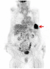
77-year-old male with colonic adenocarcinoma metastatic to the chest wall. Coronal PET image corresponding to Fig. 2 demonstrates a focus (red arrow) of high FDG uptake projecting in the lateral left basilar region, within known left basilar chest wall mass consistent with metastatic disease. Maximum SUV was 1.9.
Figure 4
Figure 4
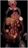
77-year-old male with colonic adenocarcinoma metastatic to the chest wall. Fused PET/CT image corresponding to Fig. 2 confirms location and exophytic morphology of a left lower chest wall mass with maximum SUV of 1.9.
Figure 5
Figure 5
77-year-old male with colonic adenocarcinoma metastatic to the chest wall. Third day of admission non-contrast axial 5 mm CT image (window level 40, window width 400, 120 kV, DLP 477, Siemens Sensation 40) of the lower chest shows an exuberate large lobulated soft tissue density chest wall mass with evidence of rib destruction anteriorly. Minimal left pleural fluid layers dependently. Average attenuation is 38.3 HU, with standard deviation = 24.4, consistent with acute bleeding.
Figure 6
Figure 6
77-year-old male with colonic adenocarcinoma metastatic to the chest wall. Eleventh day of admission high resolution axial CT image (window level 400, window width 350, 120 kV, DLP 614, Siemens Definition AS+) following administration of intravenous contrast and drainage procedure shows marked decrease in size of the chest wall mass (red arrow) with a residual soft tissue density mass centered about the left anterior 7th rib, evidence of rib destruction and subcutaneous emphysema. The lesion’s mean attenuation is 28.8 HU with standard deviation 131.6.
Figure 7
Figure 7
77-year-old male with colonic adenocarcinoma metastatic to the chest wall. Low magnification (40X) view of chest wall biopsy specimen shows a moderately differentiated adenocarcinoma infiltrating bone and fibroconnective tissue. The glands are lined by columnar cells with peripheral palisading and show central dirty necrosis, characteristic of colonic adenocarcinoma. This morphology is similar to that of the 2007 resection of metastasis to lung.
Figure 8
Figure 8
77-year-old male with colonic adenocarcinoma metastatic to the chest wall. High magnification (100X) view of chest wall biopsy specimen shows a moderately differentiated adenocarcinoma infiltrating bone and fibroconnective tissue. The glands are lined by columnar cells with peripheral palisading and show central dirty necrosis, characteristic of colonic adenocarcinoma. This morphology is similar to that of the 2007 resection of metastasis to lung.

References

    1. Ferlay J, Shin HR, Bray F, Forman D, Mathers C, Parkin DM. Estimates of worldwide burden of cancer in 2008: GLOBOCAN 2008. International Journal of Cancer. 2010;127:2893–2917. - PubMed
    1. Ko FC, Liu JM, Chen WS, Chiang JK, Lin TC, Lin JK. Risk and patterns of brain metastases in colorectal cancer: 27-year experience. Dis Colon Rectum. 1999;42:1467–1471. - PubMed
    1. O’Sullivan P, O’Dwyer H, Flint J, Munk PL, Muller N. Soft tissue tumours and mass-like lesions of the chest wall: a pictorial review of CT and MR findings. Br J Radiol. 2007;80(955):574–80. - PubMed
    1. Hong WK. Chest Wall Tumors. Merck manual online. Retrieved from http://http://www.merckmanuals.com/professional/pulmonary_disorders/tumo....
    1. Jiang L, Gao Y, Sheng S, Xu M, Lu L, Lu H. A first described chest wall metastasis from colon cancer demonstrated with (18)F-FDG PET/CT. Hell J Nucl Med. 2011;14(3):316–7. - PubMed

Publication types

LinkOut - more resources