

A case of malignant peritoneal mesothelioma revealed with limitation of PET-CT in the diagnosis of thoracic metastasis
- PMID: 23372960
- PMCID: PMC3547994
- DOI: 10.3978/j.issn.2072-1439.2012.08.19
A case of malignant peritoneal mesothelioma revealed with limitation of PET-CT in the diagnosis of thoracic metastasis
Abstract
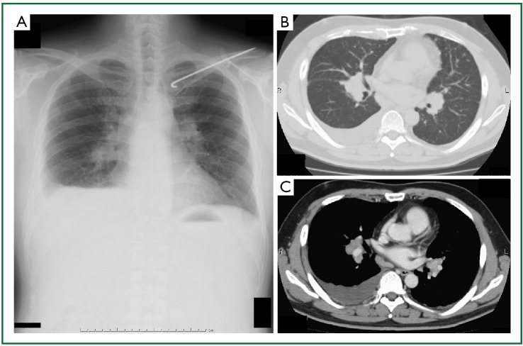
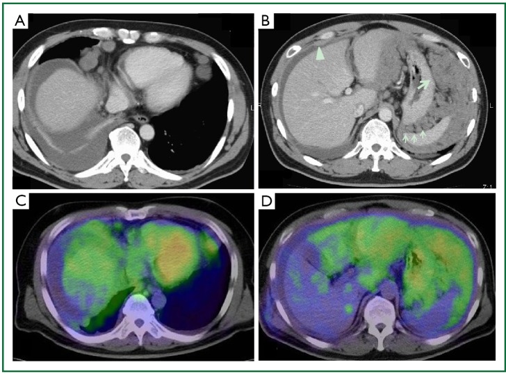
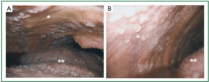
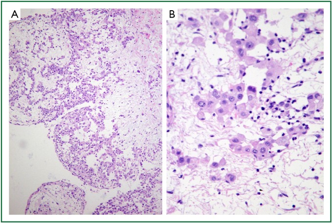
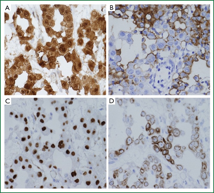
A 47-year-old man was referred to our hospital because of a 2-month history of dry cough, 2-kg weight loss, and a feeling of abdominal fullness. The PET-CT scan depicts the intense standard uptake values (SUVs) of the anterior and subphrenic lymphnodes, and intraperitoneal cavity, especially in the omentum, while, no uptake was found in the pleural cavity. Based on the pathological findings of the open lung biopsy specimens, he was diagnosed with malignant peritoneal mesothelioma of epithelioid type with thoracic metastasis. The present case demonstrated the some of the limitations of PET-CT in the diagnosis of malignant mesothelioma, which failed to detect pleural involvement despite aggressive invasion by this tumor.
Keywords: Malignant peritoneal mesothelioma; fluorodeoxyglucose (FDG) positron emission tomography (PET) computed tomography; open lung biopsy; thoracic metastasis.
Figures
Similar articles
-
A Case of Localized Malignant Peritoneal Mesothelioma Evaluated by 18F-FDG PET/CT.Clin Nucl Med. 2020 Nov;45(11):890-891. doi: 10.1097/RLU.0000000000003158. Clin Nucl Med. 2020. PMID: 32604114
-
18F-FDG PET/CT in a recurrent diffuse malignant peritoneal mesothelioma.Clin Nucl Med. 2012 May;37(5):492-4. doi: 10.1097/RLU.0b013e3182478bb5. Clin Nucl Med. 2012. PMID: 22475902
-
Clinical implications of 18F-fluorodeoxyglucose positron emission tomography/computed tomography at delayed phase for diagnosis and prognosis of malignant pleural mesothelioma.Oncol Rep. 2012 Feb;27(2):333-8. doi: 10.3892/or.2011.1520. Epub 2011 Oct 24. Oncol Rep. 2012. PMID: 22024889
-
Does positron emission tomography offer prognostic information in malignant pleural mesothelioma?Interact Cardiovasc Thorac Surg. 2011 May;12(5):806-11. doi: 10.1510/icvts.2010.255901. Epub 2011 Jan 25. Interact Cardiovasc Thorac Surg. 2011. PMID: 21266493 Review.
-
What is the best way to diagnose and stage malignant pleural mesothelioma?Interact Cardiovasc Thorac Surg. 2011 Feb;12(2):254-9. doi: 10.1510/icvts.2010.255893. Epub 2010 Nov 1. Interact Cardiovasc Thorac Surg. 2011. PMID: 21044972 Review.
Cited by
-
Massive localized malignant pleural mesothelioma (LMPM): manifestations on computed tomography in 6 cases.Int J Clin Exp Med. 2015 Oct 15;8(10):18367-74. eCollection 2015. Int J Clin Exp Med. 2015. PMID: 26770440 Free PMC article.
-
Pleuroperitoneal Mesothelioma: A Rare Entity on 18F-FDG PET/CT.Indian J Nucl Med. 2017 Jan-Mar;32(1):75-76. doi: 10.4103/0972-3919.198499. Indian J Nucl Med. 2017. PMID: 28242997 Free PMC article.
-
Amelanotic Malignant Melanoma with Dense Pleural Thickening Mimicking Malignant Mesothelioma.Intern Med. 2019 Apr 1;58(7):969-972. doi: 10.2169/internalmedicine.0867-18. Epub 2018 Nov 19. Intern Med. 2019. PMID: 30449771 Free PMC article.
-
Serendipitous discovery of peritoneal mesothelioma.Proc (Bayl Univ Med Cent). 2016 Apr;29(2):185-7. doi: 10.1080/08998280.2016.11929410. Proc (Bayl Univ Med Cent). 2016. PMID: 27034564 Free PMC article.
-
Significant Clinical Benefit of Pemetrexed-based Chemotherapy for Advanced Diffuse Malignant Peritoneal Mesothelioma: A Case Presentation.Indian J Med Paediatr Oncol. 2017 Jan-Mar;38(1):73-77. doi: 10.4103/0971-5851.203495. Indian J Med Paediatr Oncol. 2017. PMID: 28469343 Free PMC article.
References
-
- Nozaki M, Suzuki T, Takahashi H, et al. A case of peritoneal malignant mesothelioma. Nihon Shokakibyo Gakkai Zasshi 2003;100:610-2 - PubMed
-
- Legha SS, Muggia FM. Pleural mesothelioma: clinical features and therapeutic implications. Ann Intern Med 1977;87:613-21 - PubMed
-
- Hillerdal G.Malignant mesothelioma 1982: review of 4710 published cases. Br J Dis Chest 1983;77:321-43 - PubMed
-
- Moertel CG. Peritoneal mesothelioma. Gastroenterology 1972;63:346-50 - PubMed
Publication types
LinkOut - more resources
Full Text Sources