

Intracranial phosphaturic mesenchymal tumor, mixed connective tissue variant presenting without oncogenic osteomalacia
- PMID: 23372968
- PMCID: PMC3551505
- DOI: 10.4103/2152-7806.104745
Intracranial phosphaturic mesenchymal tumor, mixed connective tissue variant presenting without oncogenic osteomalacia
Abstract
Background: Phosphaturic mesenchymal tumor, mixed connective tissue variant (PMTMCT) is a rare tumor typically occurring in soft tissues and bone, causing oncogenic (tumor-induced) osteomalacia (TIO) through secretion of the phosphaturic hormone, fibroblast growth factor-23 (FGF-23). Rare tumors identical to PMTMCT occur without known TIO. Intracranial localization of PMTMCT is extremely rare, with only two cases reported in the literature. We present a very unusual case of a patient with an intracranial PMTMCT that presented with neurologic changes without osteomalacia.
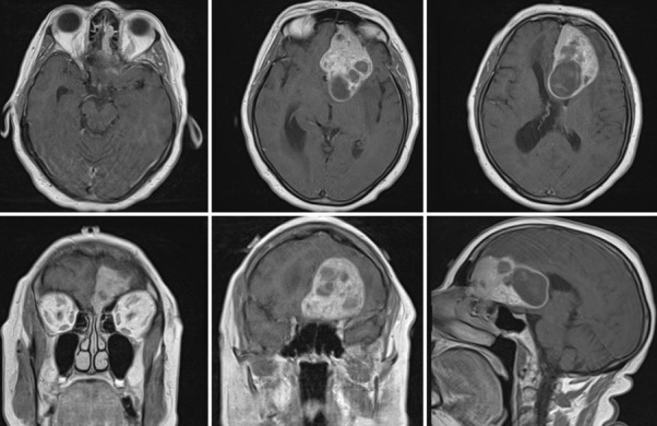
Case description: A 67-year-old woman presented with progressive incontinence, apathy, and abulia after having undergone a total knee replacement 1 month earlier. Imaging disclosed a large left frontal anterior fossa mass. She underwent uncomplicated surgical resection of this tumor. Surprisingly, histopathology suggested PMTMCT. Reverse transcription polymerase chain reaction (RT-PCR) assay demonstrating FGF-23 expression in the tumor confirmed the diagnosis. Serum FGF-23 levels postoperatively were normal and she had no clinical or laboratory evidence of osteomalacia or phosphaturia.
Conclusion: This report should serve to alert clinicians to the possibility that PMTMCT can be included in the differential diagnosis of intracranial masses even in the absence of tumor-induced osteomalacia.
Keywords: Intracranial; neoplasm; neuropathology; oncogenic osteomalacia.
Figures

References
-
- Bahrami A, Weiss SW, Montgomery E, Horvai AE, Jin L, Inwards CY, et al. RT-PCR analysis for FGF23 using paraffin sections in the diagnosis of phosphaturic mesenchymal tumors with and without known tumor induced osteomalacia. Am J Surg Pathol. 2009;33:1348–54. - PubMed
-
- Carpenter TO. Oncogenic osteomalacia-a complex dance of factors. N Engl J Med. 2003;348:1705–8. - PubMed
-
- David K, Revesz T, Kratimenos G, Krausz T, Crockard HA. Oncogenic osteomalacia associated with a meningeal phosphaturic mesenchymal tumor. Case report. J Neurosurg. 1996;84:288–92. - PubMed
-
- Evans DJ, Azzopardi JG. Distinctive tumours of bone and soft tissue causing acquired vitamin-D-resistant osteomalacia. Lancet. 1972;1:353–4. - PubMed
-
- Folpe AL, Fanburg-Smith JC, Billings SD, Bisceglia M, Bertoni F, Cho JY, et al. Most osteomalacia-associated mesenchymal tumors are a single histopathologic entity: An analysis of 32 cases and a comprehensive review of the literature. Am J Surg Pathol. 2004;28:1–30. - PubMed
Publication types
LinkOut - more resources
Full Text Sources
