. 2012:3:45.
doi: 10.4103/2153-3539.104907. Epub 2012 Dec 20.

Experience with multimodality telepathology at the University of Pittsburgh Medical Center

Affiliations

Experience with multimodality telepathology at the University of Pittsburgh Medical Center

Liron Pantanowitz et al. J Pathol Inform. 2012.

Abstract

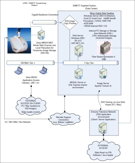
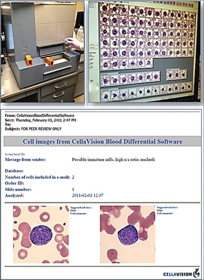
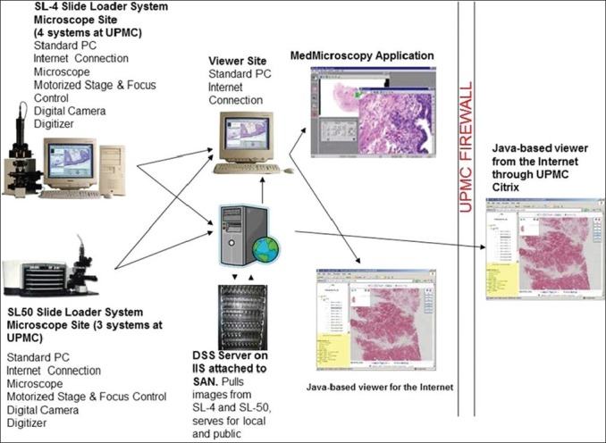
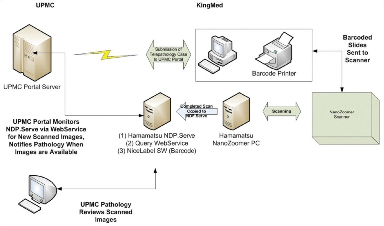
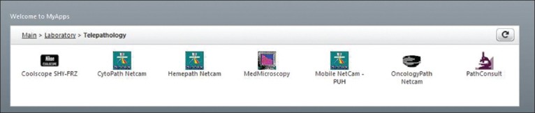
Several modes of telepathology exist including static (store-and-forward), dynamic (live video streaming or robotic microscopy), and hybrid technology involving whole slide imaging (WSI). Telepathology has been employed at the University of Pittsburgh Medical Center (UPMC) for over a decade at local, national, and international sites. All modes of telepathology have been successfully utilized to exploit our institutions subspecialty expertise and to compete for pathology services. This article discusses the experience garnered at UPMC with each of these teleconsultation methods. Static and WSI telepathology systems have been utilized for many years in transplant pathology using a private network and client-server architecture. Only minor clinically significant differences of opinion were documented. In hematopathology, the CellaVision(®) system is used to transmit, via email, static images of blood cells in peripheral blood smears for remote interpretation. While live video streaming has remained the mode of choice for providing immediate adequacy assessment of cytology specimens by telecytology, other methods such as robotic microscopy have been validated and shown to be effective. Robotic telepathology has been extensively used to remotely interpret intra-operative neuropathology consultations (frozen sections). Adoption of newer technology and increased pathologist experience has improved accuracy and deferral rates in teleneuropathology. A digital pathology consultation portal (https://pathconsult.upmc.com/) was recently created at our institution to facilitate digital pathology second opinion consults, especially for WSI. The success of this web-based tool is the ability to handle vendor agnostic, large image files of digitized slides, and ongoing user-friendly customization for clients and teleconsultants. It is evident that the practice of telepathology at our institution has evolved in concert with advances in technology and user experience. Early and continued adoption of telepathology has promoted additional digital pathology resources that are now being leveraged for other clinical, educational, and research purposes.

Keywords: Digital imaging; robotic; static; teleconsultation; telemicroscopy; telepathology; video microscopy; whole slide imaging.


Figures

Figure 1
Figure 1
Diagram showing the information technology and server components for the UPMC/ISMETT telepathology platform. Cases are accessioned in the Mirax Digital Slide Desktop (MDSD) image repository (blue box), individual slides are then scanned on the Mirax MIDI WSI scanner and transferred via network file share to the MDSD repository. Access to the images is direct via secure (username/password) connection to the MDSD system from client PC/Mac using a java applet for viewing, or via affiliated/specific workflow applications for the purpose of transplant immunology assessment. The java applet viewer can be embedded in any website or client/server application for flexibility of workflow integration
Figure 2
Figure 2
CellaVision® System. (a) CellaVision DM96 instrument; (b) Review monitor displaying captured images of different blood cells; (c) E-mail with embedded selected static images generated using the systems remote review software
Figure 3
Figure 3
Comparison between glass and telecytology using video streaming. For remote evaluations of fine needle aspirations, cytopathologists demonstrated similar performance using a conventional microscope (blue bars) and the telecytology system (yellow bars) for providing tissue adequacy (far left bars) and the correct diagnosis (far right bars). However, the middle bars in this graph indicate that pathologists found reviewing cases remotely to appear more complex, with inferior image quality and more obscured features. Consequently, they were less confident with their telecytology diagnoses
Figure 4
Figure 4
Average time (seconds) taken to review glass and telecytology cases. Cytopathologists took longer to review a cytology slide using telecytology (94 seconds) than they did when examining the same slide with a conventional light microscope (70 seconds)
Figure 5
Figure 5
Neuropathology frozen section rate (updated from Horbinski et al. 2007)[9]
Figure 6
Figure 6
Information technology infrastructure used to support intra-institutional neuropathology frozen section telepathology
Figure 7
Figure 7
Gross teleneuropathology showing close-up views of two portions of tissue submitted for frozen section from a glioblastoma multiforme tumor
Figure 8
Figure 8
Data workflow with the UPMC Portal and one of their clients (KingMed in China). The data flow utilizes a web service (deployed on the NDP. Serve server) to monitor the NDP. Serve system for completed slide scans that can be associated with a telepathology case. As scans are transferred from the scanner to the NDP. Serve database, this system allows for automated notification of the UPMC assigned pathologist that the case is ready for consult. Perhaps more importantly, this automated system eliminates a person having to email or notify, which scan filenames are associated with a case, thereby removing the possibility of human error of assigning the wrong slide image to a case
Figure 9
Figure 9
Suite of current telepathology web-based applications available for pathologist use at UPMC

References

    1. Kaplan K, Weinstein RS, Pantanowitz L. Telepathology. In: Pantanowitz L, Balis UJ, Tuthill JM, editors. Pathology Informatics: Theory and Practice. Vol. 16. Chicago: ASCP Press; 2012. pp. 257–72.
    1. Della Mea V. 25 years of telepathology research: A bibliometric analysis. Diagn Pathol. 2011;6(Suppl 1):S26. - PMC - PubMed
    1. Minervini MI, Yagi Y, Marino IR, Lawson A, Nalesnik M, Randhawa P, et al. Development and experience with an integrated system for transplantation telepathology. Hum Pathol. 2001;32:1334–43. - PubMed
    1. Isse K, Lesniak A, Grama K, Roysam B, Minervini MI, Demetris AJ. Digital transplantation pathology: Combining whole slide imaging, multiplex staining and automated image analysis. Am J Transplant. 2012;12:27–37. - PMC - PubMed
    1. Rollins-Raval MA, Raval JS, Contis L. Experience with CellaVision DM96 for peripheral blood differentials in a large multi-center academic hospital system. J Pathol Inform. 2012;3:29. - PMC - PubMed