

Posterior cranial fossa meningiomas
- PMID: 23372989
- PMCID: PMC3424023
- DOI: 10.1055/s-0032-1304835
Posterior cranial fossa meningiomas
Abstract
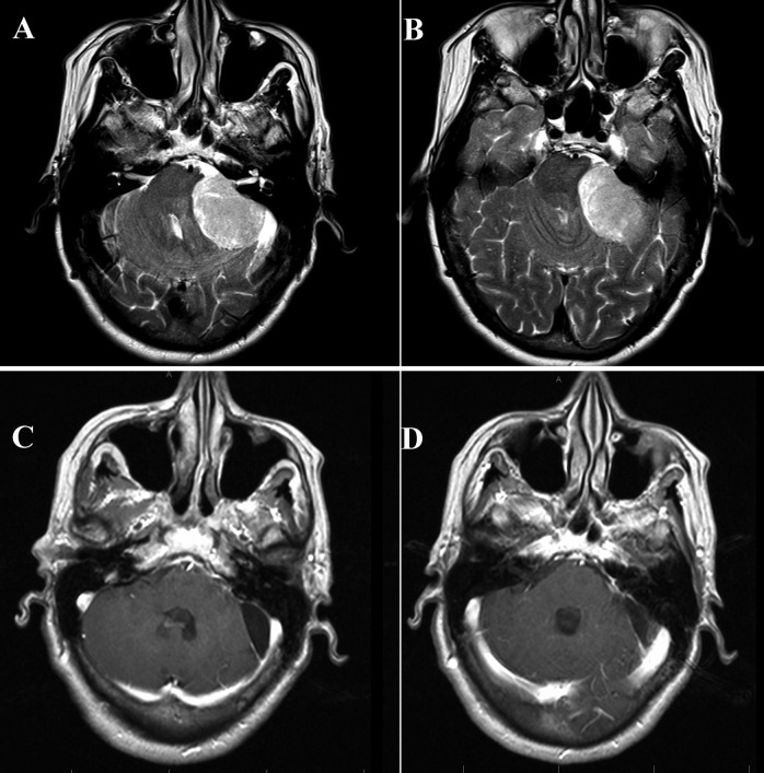
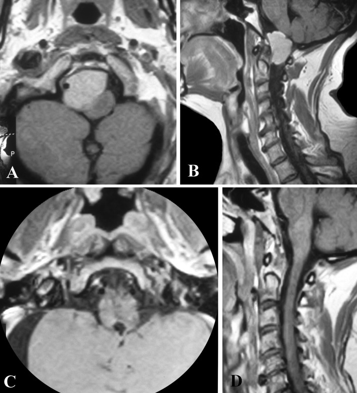
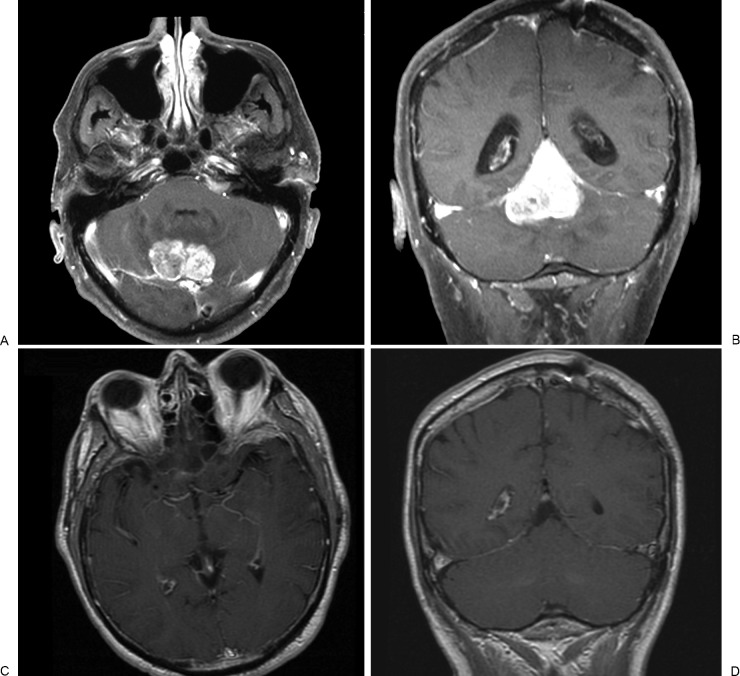
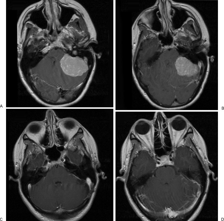
This study evaluated the outcomes, complications, and recurrence rates of posterior cranial fossa meningiomas. We retrospectively reviewed our surgical experience with 64 posterior cranial fossa meningiomas. Mean age was 56 years with a female preponderance (67.2%). Headache was the most common symptom. Retrosigmoid approach was the commonest surgical procedure (23.4%). The incidence of cranial nerve related complications was 28%. Postoperatively facial nerve weakness was observed in 11%. The incidence of cerebrospinal fluid leak was 4.6%. Gross total resection was achieved in 37 patients (58%). Sixteen patients (25%) with residual tumors underwent Gamma knife radiosurgery. Recurrence or tumor progression was observed in 12 patients (18.7%). Operative mortality was 3.1%. At their last follow-up, 93% of the cases achieved Glasgow Outcome Scale scores 4 or 5. Total excision is the ideal goal which can be achieved with meningiomas located in certain location, such as lateral convexity, but for other posterior fossa meningiomas the close proximity of critical structures is a major obstacle in achieving this goal. In practicality, a balance between good functional outcome and extent of resection is important for posterior cranial fossa meningiomas in proximity to critical structures.
Keywords: meningiomas; posterior fossa; skull base.
Figures
Similar articles
-
Petroclival meningiomas: study on outcomes, complications and recurrence rates.J Neurosurg. 2011 May;114(5):1268-77. doi: 10.3171/2010.11.JNS10326. Epub 2010 Dec 24. J Neurosurg. 2011. PMID: 21184632
-
Gamma Knife radiosurgery for posterior fossa meningiomas: a multicenter study.J Neurosurg. 2015 Jun;122(6):1479-89. doi: 10.3171/2014.10.JNS14139. Epub 2015 Apr 10. J Neurosurg. 2015. PMID: 25859812
-
Efficacy and outcomes of facial nerve-sparing treatment approach to cerebellopontine angle meningiomas.J Neurosurg. 2017 Dec;127(6):1231-1241. doi: 10.3171/2016.10.JNS161982. Epub 2017 Feb 10. J Neurosurg. 2017. PMID: 28186449
-
Meningiomas of the basal posterior fossa. Surgical experience in 80 cases.Neurocirugia (Astur). 2004 Dec;15(6):525-42. doi: 10.1016/s1130-1473(04)70439-x. Neurocirugia (Astur). 2004. PMID: 15632989 Review.
-
Meningiomas engaging major venous sinuses.World Neurosurg. 2014 Jan;81(1):116-24. doi: 10.1016/j.wneu.2013.01.095. Epub 2013 Jan 30. World Neurosurg. 2014. PMID: 23376533 Review.
Cited by
-
Surgery for posterior fossa meningioma: elevated postoperative cranial nerve morbidity discards aggressive tumor resection policy.Neurosurg Rev. 2021 Apr;44(2):953-959. doi: 10.1007/s10143-020-01275-6. Epub 2020 Feb 27. Neurosurg Rev. 2021. PMID: 32107680
-
Petroclival meningiomas: radiological features essential for surgeons.Ecancermedicalscience. 2019 Mar 5;13:907. doi: 10.3332/ecancer.2019.907. eCollection 2019. Ecancermedicalscience. 2019. PMID: 31123490 Free PMC article. Review.
-
Evaluation of Ventriculoperitoneal Shunt-Related Complications in Intracranial Meningioma with Hydrocephalus.J Neurol Surg B Skull Base. 2017 Feb;78(1):30-36. doi: 10.1055/s-0036-1584309. Epub 2016 Jun 2. J Neurol Surg B Skull Base. 2017. PMID: 28180040 Free PMC article.
-
CLINICAL AND SURGICAL CHARACTERISTICS OF POSTERIOR FOSSA TUMORS IN ADULTS - SINGLE-CENTER EXPERIENCE OF SURGICAL MANAGEMENT.Acta Clin Croat. 2023 Nov;62(3):502-509. doi: 10.20471/acc.2023.62.03.12. Acta Clin Croat. 2023. PMID: 39310684 Free PMC article.
-
Genomic Biomarkers of Meningioma: A Focused Review.Int J Mol Sci. 2021 Sep 23;22(19):10222. doi: 10.3390/ijms221910222. Int J Mol Sci. 2021. PMID: 34638590 Free PMC article. Review.
References
-
- Campbell E, Whitfield R D. Posterior fossa meningiomas. J Neurosurg. 1948;5(2):131–153. - PubMed
-
- Castellano F, Ruggiero G. Meningiomas of the posterior fossa. Acta Radiol Suppl. 1953;104:1–177. - PubMed
-
- Sekhar L N, Wright D C, Richardson R, Monacci W. Petroclival and foramen magnum meningiomas: surgical approaches and pitfalls. J Neurooncol. 1996;29(3):249–259. - PubMed
-
- Al-Mefty O, Fox J L, Smith R R. Petrosal approach for petroclival meningiomas. Neurosurgery. 1988;22(3):510–517. - PubMed
-
- Samii M, Ammirati M, Mahran A, Bini W, Sepehrnia A. Surgery of petroclival meningiomas: report of 24 cases. Neurosurgery. 1989;24(1):12–17. - PubMed
LinkOut - more resources
Full Text Sources
