

Real time parallel intraoperative integration of endoscopic, microscopic, and navigation images: a proof of concept based on laboratory dissections
- PMID: 23372993
- PMCID: PMC3424027
- DOI: 10.1055/s-0032-1304554
Real time parallel intraoperative integration of endoscopic, microscopic, and navigation images: a proof of concept based on laboratory dissections
Abstract
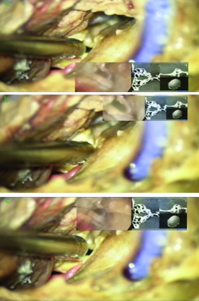
Endoscope, microscope, and neuronavigation systems are integrated in neurosurgical procedures mainly by using a serial combination algorithm, where, the user must switch his/her field of view from one platform display to another. The integration of theses devices could be optimized by incorporating different displays into one viewing platform thus achieving a parallel combination. In this study, we investigated the feasibility and the applicability of parallel integration of microscopic, endoscopic, and neuronavigation images by real time displaying the endoscope and neuronavigation image datasets in the main operative microscope oculars. The proposed set-up was effective in displaying the three images dataset in an operationally actionable mode. Ergonomically, the ability of using the different image dataset without the need of taking the eyes off the microscope oculars did not disrupt the flow or the tempo of the operative procedure. However, new endoscopes specific to this application are recommended.
Keywords: endoscope; image-guided surgery; medical technology; microneurosurgery; skull base surgery.
Figures


Similar articles
-
Endoscope-assisted brain surgery: part 1--evolution, basic concept, and current technique.Neurosurgery. 1998 Feb;42(2):219-24; discussion 224-5. doi: 10.1097/00006123-199802000-00001. Neurosurgery. 1998. PMID: 9482171
-
Multiple brain tumor nodule resections under direct visualization of a neuronavigated endoscope.Minim Invasive Neurosurg. 2007 Aug;50(4):227-32. doi: 10.1055/s-2007-985861. Minim Invasive Neurosurg. 2007. PMID: 17948182
-
Impact of a self-developed planning and self-constructed navigation system on skull base surgery: 10 years experience.Acta Otolaryngol. 2007 Apr;127(4):403-7. doi: 10.1080/00016480601002104. Acta Otolaryngol. 2007. PMID: 17453461
-
Future perspectives for intraoperative MRI.Neurosurg Clin N Am. 2005 Jan;16(1):201-13. doi: 10.1016/j.nec.2004.07.011. Neurosurg Clin N Am. 2005. PMID: 15561539 Review.
-
[Intraoperative navigation, with focus on the skull base].HNO. 2016 Sep;64(9):635-40. doi: 10.1007/s00106-016-0215-x. HNO. 2016. PMID: 27566369 Review. German.
Cited by
-
Recurrent atlantoaxial synovial cyst resection via a navigation-guided, endoscope-assisted posterior approach.Surg Neurol Int. 2014 Dec 30;5(Suppl 15):S567-9. doi: 10.4103/2152-7806.148048. eCollection 2014. Surg Neurol Int. 2014. PMID: 25593779 Free PMC article.
-
Frameless Stereotactic Insertion of Viewsite Brain Access System with Microscope-Mounted Tracking Device for Resection of Deep Brain Lesions: Technical Report.Cureus. 2017 Feb 4;9(2):e1012. doi: 10.7759/cureus.1012. Cureus. 2017. PMID: 28331774 Free PMC article.
-
Minimalistic Approaches to Craniovertebral Junction Tumors.Adv Tech Stand Neurosurg. 2025;55:165-179. doi: 10.1007/978-3-031-90762-3_9. Adv Tech Stand Neurosurg. 2025. PMID: 40608106
References
-
- Cappabianca P, Cinalli G, Gangemi M. et al.Application of neuroendoscopy to intraventricular lesions. Neurosurgery. 2008;62(Suppl 02):575–597, discussion 597–598. - PubMed
-
- Di Rocco F, Oi S, Samii A. et al.Neuronavigational endoscopic endonasal sellar and parasellar surgery using a 2-mm-diameter lens rigid-rod endoscope: a cadaver study. Neurosurgery. 2007;60(4, Suppl 02):394–400, discussion 400. - PubMed
-
- Eloy J A, Carai A, Patel A B, Genden E M, Bederson J B. Combined endoscope-assisted transclival clipping and endovascular stenting of a basilar trunk aneurysm: case report. Neurosurgery. 2008;62(3, Suppl 01):142–143, discussion 143–144. - PubMed
-
- Greenfield J P, Leng L Z, Chaudhry U. et al.Combined simultaneous endoscopic transsphenoidal and endoscopic transventricular resection of a giant pituitary macroadenoma. Minim Invasive Neurosurg. 2008;51(5):306–309. - PubMed
-
- Rhoten R L, Luciano M G, Barnett G H. Computer-assisted endoscopy for neurosurgical procedures: technical note. Neurosurgery. 1997;40(3):632–637, discussion 638. - PubMed
LinkOut - more resources
Full Text Sources
Other Literature Sources
