. 2013 Mar 26;7(3):2032-41.
doi: 10.1021/nn305962n. Epub 2013 Feb 7.

Fluorochrome-functionalized nanoparticles for imaging DNA in biological systems

Affiliations

Fluorochrome-functionalized nanoparticles for imaging DNA in biological systems

Hoonsung Cho et al. ACS Nano. .

Abstract

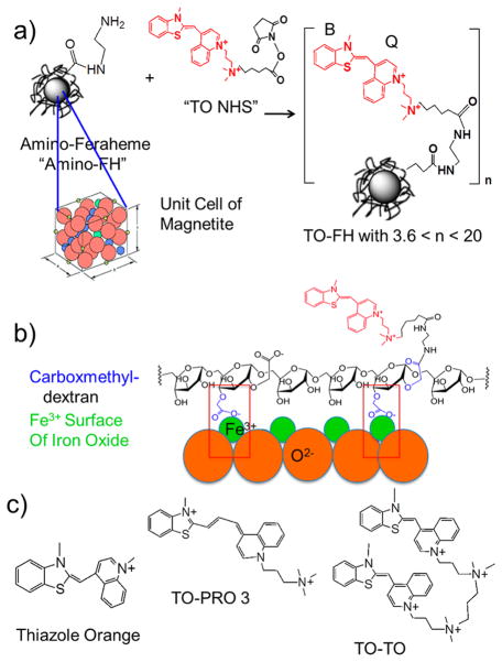
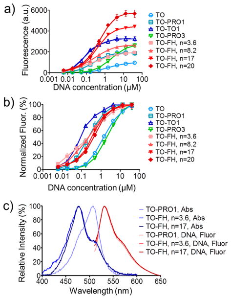
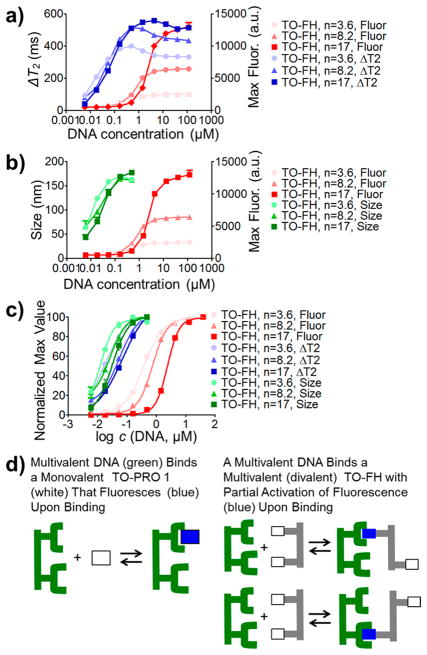
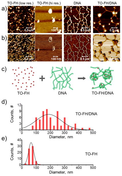
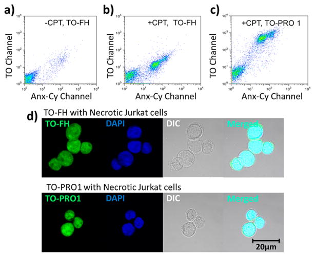
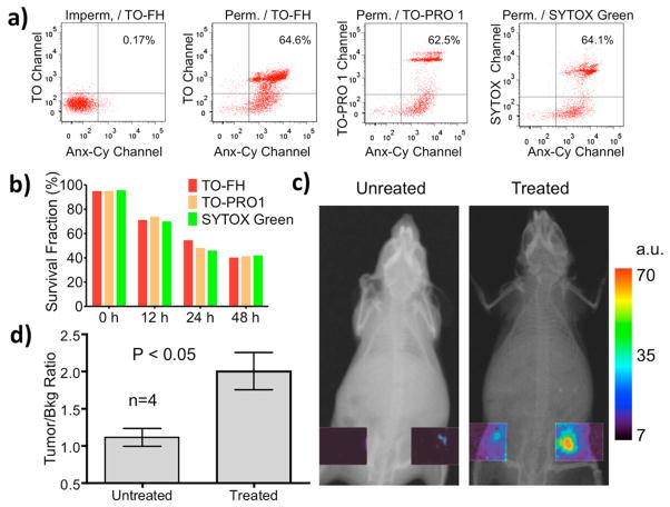
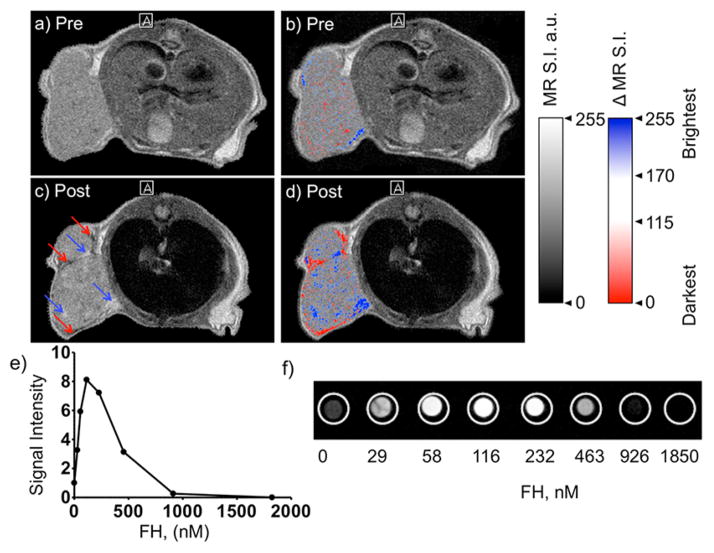
Attaching DNA binding fluorochromes to nanoparticles (NPs) provides a way of obtaining NPs that bind to DNA through fluorochrome mediated interactions. To obtain a nanoparticle (NP) that bound to the DNA in biological systems, we attached the DNA binding fluorochrome, TO-PRO 1 (TO), to the surface of the Feraheme (FH) NP, to obtain a fluorochrome-functionalized NP denoted TO-FH. When reacted with DNA in vitro, TO-FH formed microaggregates that were characterized by fluorescence, light scattering, and T2 changes. The formation of DNA/TO-FH microaggregates was also characterized by AFM, with microaggregates exhibiting a median size of 200 nm, and consisting of DNA and multiple TO-FH NPs whose individual diameters were only 25-35 nm. TO-FH failed to bind normal cells in culture, but treatment with chemotherapeutic agents or detergents yielded necrotic cells that bound TO-FH and vital fluorochromes similarly. The uptake of TO-FH by HT-29 xenografts (treated with 5-FU and oxaliplatin) was evident by surface fluorescence and MRI. Attaching multiple DNA binding fluorochromes to magnetic nanoparticles provides a way of generating DNA binding NPs that can be used to detect DNA detection by microaggregate formation in vitro, for imaging the DNA of necrotic cells in culture, and for imaging the DNA of a tumor treated with a chemotherapeutic agent. Fluorochrome functionalized NPs are a multimodal (magnetic and fluorescent), highly multivalent (n ≈ 10 fluorochromes/NP) nanomaterials useful for imaging the DNA of biological systems.


Conflict of interest statement

Conflict of Interest: The authors declare no competing financial interest.

Figures

Figure 1
Figure 1
Synthesis and properties of fluorochrome-functionalized NPs. (a) Reacting the “TO NHS ester” with the amino-Feraheme generated fluorochrome-functionalized NPs bearing different numbers of fluorochromes (n) per NP. The benzothiazole (B) and quinoline (Q) rings of TO-PRO1 are shared by the low molecular reference fluorochromes: thiazole orange, TO-PRO 3, and TO-TO whose structures are given in Figure 1c. (b) Surface chemistry for fluorochrome-functionalized NPs. The carboxymethyldextran provides carboxyl groups for fluorochrome attachment and bind to the iron oxide, generating high thermal stability. (c) Structures of reference fluorochromes containing the benzothiazole (B) and quinoline (Q) rings of TO-PRO 1. Structure of TO-PRO 1 is in red in panel A.
Figure 2
Figure 2
Reaction between fluorochrome-functionalized NPs, with different numbers of TO-PRO 1 per NP (n), and DNA as obtained by fluorescence. Data were obtained at 50 nM in benzothiazole equivalents with variable NP concentrations. Fluorescence responses from panel a were fit to the logit equation as shown in panel b, with the EC50 and maximum fluorescence values (max. fluor) given in Table 2. Thiazole orange and TO-PRO 3 had higher EC50 values, while fluorochrome-functionalized NPs had similar EC50 values. (c) Absorption and emission spectra of TO-FH NPs (n = 3.6 or 17) and TO-PRO 1. TO-FH spectra differ from TO-PRO 1 spectra due to fluorochrome/fluorochrome stacking interactions on the NP surface.
Figure 3
Figure 3
Reaction between TO-FHs with DNA by techniques that measure NP/DNA microaggregate formation. Microaggregate response (at a constant NP concentration of 15.2 nM) was measured by T2 changes (a) or by light scattering (b). Fluorescence response, which differed from that of Figure 2 because of the use of fixed NP concentrations, is also shown in panels a and b. Data were analyzed by the logit equation (c) with values provided in Table 2. (d) Monovalent and divalent binding modes: the monomeric TO-PRO 1 produces its full fluorescence (blue) upon binding to DNA. A multivalent, fluorochrome-functionalized NP, represented as simplified divalent fluorochrome, has binding modes that are inefficient with respect to fluorochrome activation which occurs on DNA binding (blue).
Figure 4
Figure 4
AFM images of DNA/TO-FH microaggregates and TO-FH. Topographic (a) and phase images (b) of DNA/TO-FH microaggregates, DNA, and TO-FH. (c) Schematic of the binding to TO-FH to DNA; (d) size distribution of DNA/TO-FH microaggregates; (e) size distribution of TO-FH. Lines are Gaussian distributions.
Figure 5
Figure 5
Interaction of TO-FH or TO-PRO 1 with the DNA of Jurkat T cells. Cells were induced into necrosis by camptothecin (CPT). (a) Dual wavelength scatter plots for control cells (−CPT) reacted with an annexin V-Cy5.5 (Anx-Cy) and TO-FH are shown. With −CPT, a small Anx-Cy, TO-FH positive population is obtained. Scatter plots for CPT-treated cells reacted with Anx-Cy and TO-FH (b) or reacted with Anx-Cy and TO-PRO 1 (c) are shown. CPT increased the population of necrotic cells that bound both TO-FH and Anx-Cy or both TO-PRO 1 and Anx-Cy. (d) Confocal microscopy of CPT treat cells reacted with DAPI and TO-FH or with DAPI and TO-PRO 1. TO-FH bound the DNA of CPT treated (but not normal) Jurkat cells by FACS or confocal microscopy.
Figure 6
Figure 6
Interaction of TO-FH with HT-29 cells after permeabilization or after exposure to 5-FU/oxaliplatin treatment to induce cell death. (a) Normal or permeabilized cells were reacted with Anx-Cy and TO-FH or TO-PRO 1 or Sytox Green. (b) Cells were treated with 5-FU/oxaliplatin and exposed to Anx-Cy plus the indicated fluorochrome. Data are plotted as the survival fraction versus time of expose. Survival fraction is the percent of cells failing to bind both Anx-Cy and a second fluorochrome, i.e., the lower left-hand quadrant of the scatter plot. With an increasing duration of treatment, the survival fraction falls. Survival fraction falls similarly with Anx-Cy and any of the three vital fluorochromes, TO-FH, TO-PRO 1, Sytox Green. (c) Tumor surface fluorescence after TO-FH injection with untreated and treated (5-FU/oxaliplatin) HT-29 xenografts. (d) Tumor fluorescence, measured as tumor/bkg fluorescence (p < 0.05), n = 4.
Figure 7
Figure 7
MR imaging of TO-FH uptake by the HT-29 tumor treated with oxaliplatin and 5-FU. (a) Pre-TO-FH image and (b) image from panel a with areas of highest signal intensity (brightest, blue) and lowest signal intensity (darkest, red) shown. Tumor is relatively uniform in signal intensity by MR signal intensity (a), with few areas high or low signal intensity shown by colorization (b). (c) Postinjection MR image. Regions of brightening (blue arrows) and darkening (red arrows) are seen. (d) Image from panel c with areas of highest signal intensity (brightest, blue) and lowest signal intensity (darkest, red) shown. (e) Signal intensity of FH solutions at varying concentrations of FH, in nM FH crystal, are shown from images (f). Using a T1 weighted pulse sequence, TO-FH can brighten or darken MR images, either of a tumor (d) or phantoms (e, f).

Similar articles

Cited by

References

    1. Marko MA, Chipperfield R, Birnboim HC. A Procedure for the Large-Scale Isolation of Highly Purified Plasmid DNA Using Alkaline Extraction and Binding to Glass Powder. Anal Biochem. 1982;121:382–387. - PubMed
    1. Boom R, Sol CJ, Salimans MM, Jansen CL, Wertheimvan Dillen PM, van der Noordaa J. Rapid and Simple Method for Purification of Nucleic Acids. J Clin Microbiol. 1990;28:495–503. - PMC - PubMed
    1. Ito T, Smith CL, Cantor CR, Sequence-Specific DNA. Purification by Triplex Affinity Capture. Proc Natl Acad Sci US A. 1992;89:495–498. - PMC - PubMed
    1. Rosi NL, Giljohann DA, Thaxton CS, Lytton-Jean AK, Han MS, Mirkin CA. Oligonucleotide-Modified Gold Nanoparticles for Intracellular Gene Regulation. Science. 2006;312:1027–1030. - PubMed
    1. Josephson L, Perez JM, Weissleder RW. Magnetic Nanosensors for the Detection of Oligonucleotide Sequences. Angew Chem Int Ed. 2001;40:3204–3206. - PubMed

MeSH terms