. 2013 Apr;125(2):193-204.
doi: 10.1111/jnc.12178. Epub 2013 Feb 27.

Dopamine D2 receptor activation leads to an up-regulation of glial cell line-derived neurotrophic factor via Gβγ-Erk1/2-dependent induction of Zif268

Affiliations

Dopamine D2 receptor activation leads to an up-regulation of glial cell line-derived neurotrophic factor via Gβγ-Erk1/2-dependent induction of Zif268

Somayeh Ahmadiantehrani et al. J Neurochem. 2013 Apr.

Abstract

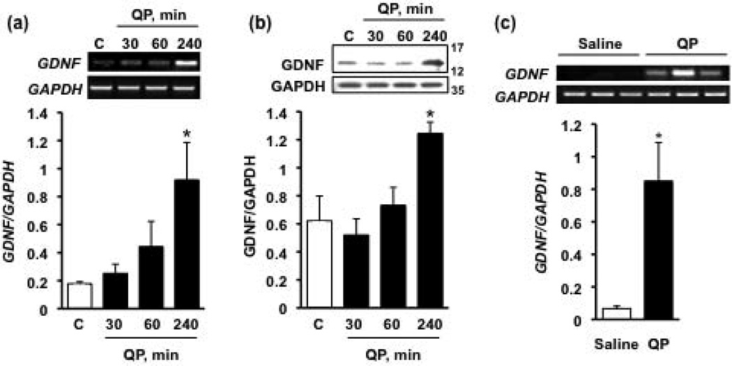
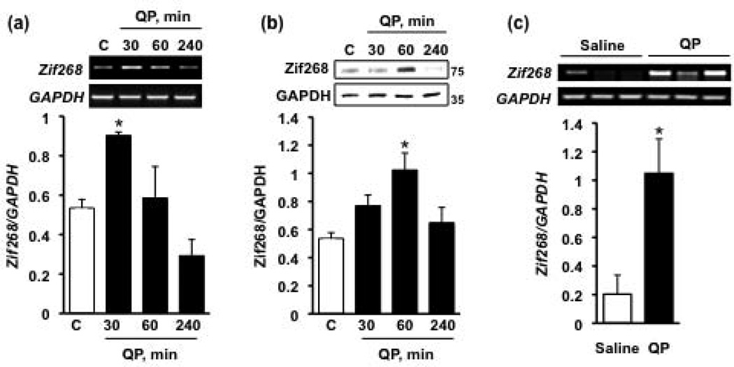
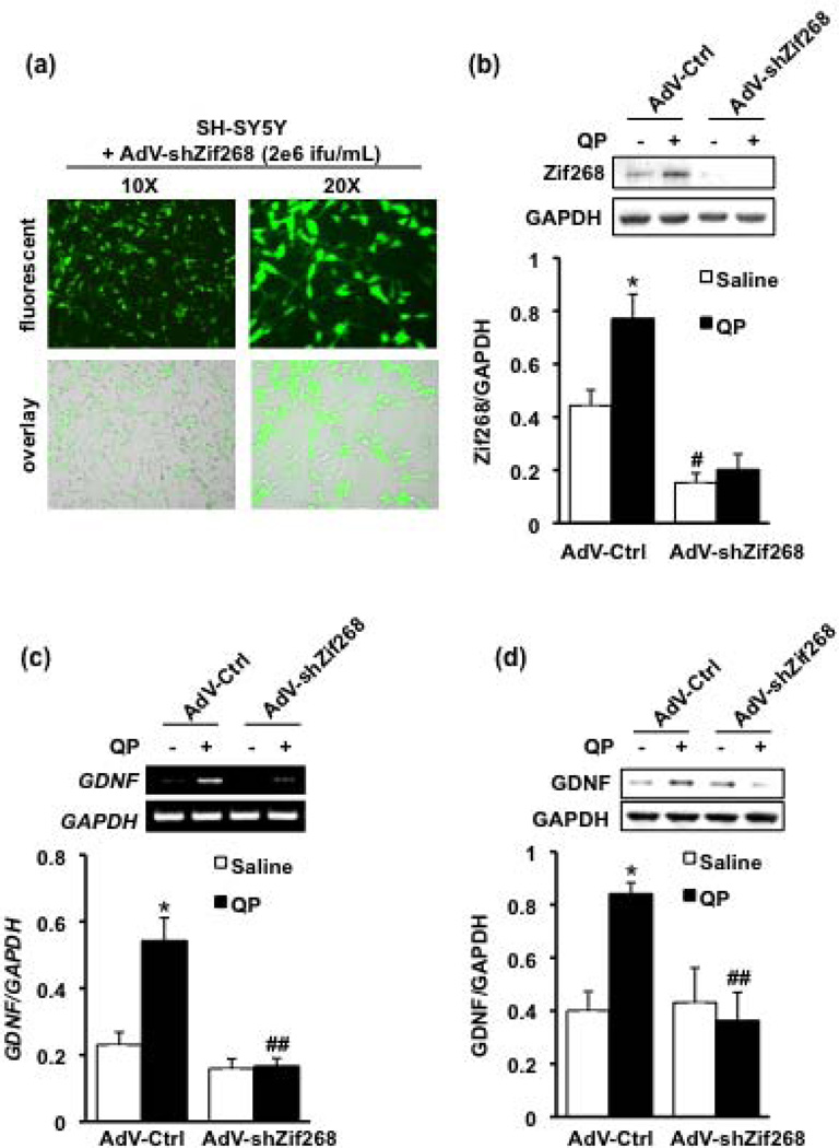
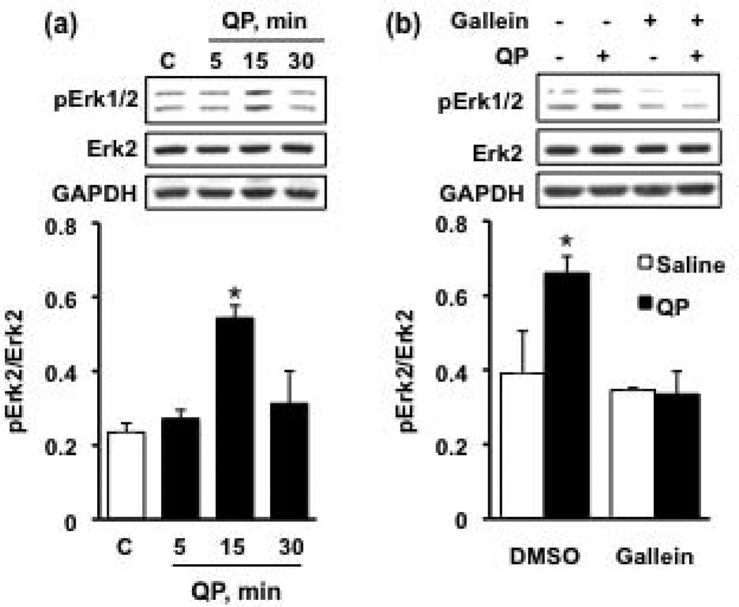
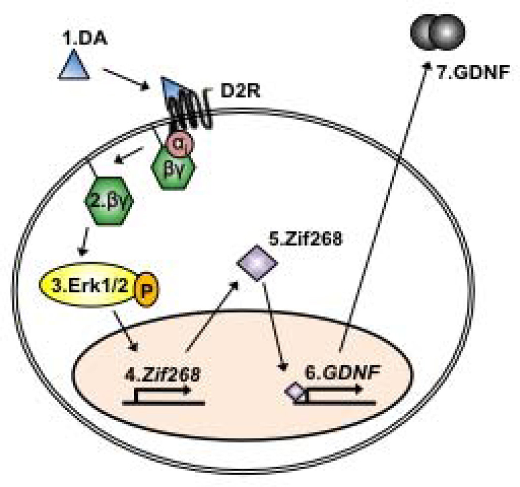
Glial cell line-derived neurotrophic factor (GDNF) is a potent growth factor essential to the development, survival, and function of dopaminergic neurons (Airaksinen and Saarma 2002). The molecular mechanisms underlying GDNF expression remain elusive; thus, we set out to identify a signaling pathway that governs GDNF levels. We found that treatment of both differentiated dopaminergic-like SH-SY5Y cells and rat midbrain slices with the dopamine D2 receptor (D2R) agonist, quinpirole, triggered an increase in the expression of GDNF that was temporally preceded by an increase in the levels of zinc-finger protein 268 (Zif268), a DNA-binding transcription factor encoded by an immediate-early gene. Moreover, the D2R inhibitor raclopride blocked the increase of both GDNF and Zif268 expression following potassium-evoked dopamine release in SH-SY5Y cells. We used adenoviral delivery of small hairpin RNA (shRNA) targeting Zif268 to down-regulate its expression and found that Zif268 is specifically required for the D2R-mediated up-regulation of GDNF. Furthermore, the D2R-mediated induction of GDNF and Zif268 expression was dependent on Gβγ-mediated signaling and activation of extracellular signal-regulated kinase 1/2. Importantly, using chromatin immunoprecipitation assay, we identified a direct association of Zif268 with the GDNF promoter. These results suggest that D2R activation induces a Gβγ- and extracellular signal-regulated kinase 1/2-dependent increase in the level of Zif268, which functions to directly up-regulate the expression of GDNF.

Keywords: Dopamine D2 receptor; Erk1/2; GDNF; Gβγ; Zif268.


Conflict of interest statement

The authors have no conflict of interest to declare.

Figures

Figure 1
Figure 1. D2R activation increases GDNF mRNA and protein levels
Cells were treated with the D2R agonist quinpirole (QP, 50 µM) for 30, 60 or 240 minutes (black bars) or saline (white bars). RT-PCR was used to assess the mRNA levels of GDNF (a) and western blot analysis was used to measure protein levels of GDNF (b). Images are representative of 4 independent experiments. Bar graphs depict the mean GDNF mRNA or protein levels (upper panels of images) normalized to the housekeeping gene GAPDH (lower panels of images) ± SEM. *p < 0.05, as compared with saline (one-way ANOVA with post hoc Student-Newman-Keuls).(c) Rat ventral midbrain slices were treated with 50 µM QP (black bar) or saline (white bar) for 240 minutes, and GDNF mRNA levels were determined via RT-PCR. *p < 0.05, as compared with saline (Student’s t-test); n = 3 samples per treatment (2 animals per sample).
Figure 2
Figure 2. D2R activation induces a rapid upregulation of Zif268
Cells were treated with the D2R agonist quinpirole (QP, 50 µM) for 30, 60 or 240 minutes (black bars) or saline (white bars). RT-PCR was used to assess the mRNA levels of Zif268 (a), while western blot analysis was used to measure protein levels of the transcription factor (b). Images are representative of 4 independent experiments. Bar graphs depict the mean Zif268 mRNA or protein levels (upper panels of images) normalized to GAPDH (lower panels of images) ± SEM. *p < 0.05, as compared with saline-treated cells (one-way ANOVA with post hoc Student-Newman-Keuls). (c) Rat ventral midbrain slices were treated with 50 µM QP (black bar) or saline (white bar) for 30 minutes, and Zif268 mRNA levels were determined via RT-PCR. *p < 0.05, as compared with saline (Student’s t-test); n = 3 samples per treatment (2 animals per sample).
Figure 3
Figure 3. Dopamine-mediated upregulation of Zif268 and GDNF is D2R-dependent
Dopamine release from cells was evoked with KCl (100 mM, final concentration) in the presence of either saline (white bars) or 10 µM Raclopride (black bars). (a) Levels of Zif268 and GDNF mRNA were determined via RT-PCR 30 and 240 minutes after the addition of KCl. Image is representative of 3 independent experiments. Bar graphs depict the mean Zif268 (b) or GDNF (c) mRNA (upper and middle panels in (a), respectively) normalized to the housekeeping gene, GAPDH (lower panel in (a)) ± SEM. *p < 0.05, as compared with no KCl, and **#p < 0.05, as compared with Saline (two-way ANOVA with post hoc Student-Newman-Keuls pair-wise comparisons).
Figure 4
Figure 4. Adenoviral-mediated knockdown of Zif268 attenuates the D2R-induced upregulation of GDNF
(a & b) Cells were infected with adenovirus expressing shRNA targeting Zif268 (AdV-shZif268), or a non-related control sequence (AdV-Ctrl), at a concentration of 2 × 10⁶ infectious units (ifu)/ml. (a) Viral infection of the SH-SY5Y cells was confirmed by GFP fluorescence 48 hours after the addition of the virus to the cell medium. (b) Cells were then treated with quinpirole (QP, 50 µM) for 60 minutes (black bars), or saline (white bars), and western blot analysis was conducted to assess Zif268 protein levels. Image is representative of 4 independent experiments. Bar graph depicts the mean Zif268 protein (upper panel) normalized to the housekeeping gene GAPDH (lower panel) ± SEM. *p < 0.05, as compared with saline, and #p < 0.05, as compared with AdV-Ctrl (two-way ANOVA with post hoc Student-Newman-Keuls pair-wise comparisons). (c & d) Cells were infected with AdV-shZif268 or AdV-Ctrl, and treated 48 hours later with 50 uM quinpirole (QP) for 240 minutes (black bars) or saline (white bars). Control groups were treated with saline. (c) RT-PCR analysis was used to assess GDNF expression. Image is representative of 4 independent experiments. (d) GDNF protein levels were evaluated by western blot. Image is a representative of 4 independent experiments. Bar graphs depict the mean GDNF mRNA (c) or protein (d) (upper panels of images) normalized to GAPDH (lower panels of images) ± SEM. *p < 0.05 as compared with the saline-treated, AdV-Ctrl group; ##p < 0.01 as compared with the quinpirole-treated, AdV-Ctrl group (two-way ANOVA with post hoc Student-Newman-Keuls pair-wise comparisons).
Figure 5
Figure 5. Activation of the D2R in SH-SY5Y cells results in a Gβγ-dependent increase in Erk1/2 phosphorylation
(a) Cells were treated with quinpirole (QP, 50µM) for 5, 15, and 30 minutes (black bars) or saline (white bar). Western blot was used to measure levels of phosphorylated Erk1/2 (pErk1/2). Image is a representative of 4 independent experiments. Levels of pErk2 (lower band in the upper panel) were normalized to the total Erk2 protein levels. GAPDH was used as a loading control. Bar graph represents the mean immunoreactivity signal of pErk2/Erk2 ± SEM. *p < 0.05, as compared with saline (one-way ANOVA with post-hoc Student-Newman-Keuls). (b) Cells pretreated for 10 minutes with the Gβγ inhibitor gallein (20µM, in DMSO vehicle) or an equal volume of DMSO at a final concentration of 0.1%. Cells were then treated with 50 µM quinpirole (black bars) or saline (white bars) for 15 minutes, the timepoint of maximal Erk1/2 pathway activation determined in (a). Western blot analysis was used to assess the level of pErk1/2, as in (a). Image is a representative of 4 independent experiments. Bar graph represents the mean immunoreactivity signal of pErk2/Erk2 ± SEM. *p < 0.05, as compared with the saline-treated control (two-way ANOVA with post-hoc Bonferroni comparisons within each pretreatment).
Figure 6
Figure 6. D2R-mediated upregulation of Zif268 and GDNF is Gβγ- and Erk1/2-dependent
Cells were pretreated for 10 minutes with the Gβγ inhibitor gallein (20 µM, in DMSO vehicle), or the MEK inhibitors U0126 or PD98059 (both at a concentration of 10 µM, in DMSO vehicle), or an equal volume of DMSO at a final concentration of 0.1%. Cells were then incubated for 30 minutes (a) or 240 minutes (b) with quinpirole (QP; 50 µM; black bars) or saline (white bars). The level of Zif268 (a) and GDNF (b) expression was determined by RT-PCR. Images are representative of 4 independent experiments. Zif268 and GDNF levels (upper panels) were normalized to those of GAPDH (lower panels). Bar graphs depict the mean Zif268 or GDNF/GAPDH ± SEM (white bars: saline-treated groups; black bars: QP-treated groups). ***p < 0.001, as compared with the saline-treated control (two-way ANOVA with post-hoc Bonferroni comparisons within each pretreatment).
Figure 7
Figure 7. D2R activation results in direct association of Zif268 at a novel Zif268 binding sequence within the GDNF promoter
(a) Schematic representation of the human GDNF promoter region 1000 base pairs upstream of the transcription start site (designated as +1). ChIP PCR products for putative Zif268 binding sites #1 (PCR1) and #2 (PCR2), as well as an intermediate region not expected to associate with Zif268 (PCR3) are depicted with solid lines. Dashed arrows represent the primers used for each PCR reaction. (b) Cells were treated with 50 µM quinpirole (QP) or saline for 60 minutes, followed by ChIP using rabbit anti-Zif268 antibodies, or normal rabbit IgG, as a control. PCRs were conducted with the resulting precipitated DNA. As a positive control (input), total DNA was used. Image is representative of 3 independent ChIP experiments.
Figure 8
Figure 8. Model: Dopamine-mediated upregulation of GDNF expression occurs via a D2R-Gβγ-Erk1/2-Zif268 pathway
Dopamine (DA) binds to and activates D2R (1), leading to the dissociation of the of the Gαi subunit from Gβγ (2). Gβγ mediates the activation of MEK and the subsequent activation of Erk1/2 (3). D2R-Gβγ-mediated activation of Erk12 results in an upregulation of Zif268 mRNA (4), which translates to an increase in the protein levels of this transcription factor (5). Zif268 directly binds the GDNF promoter region (6), and specifically promotes the transcription of GDNF, leading to an increase in the levels of GDNF mRNA and protein (7).

References

    1. Ahmadiantehrani S, Barak S, Ron D. GDNF is a novel ethanol-responsive gene in the VTA: implications for the development and persistence of excessive drinking. Addict Biol. 2013 In press. - PMC - PubMed
    1. Airaksinen M, Saarma M. The GDNF family: signaling, biological functions and therapeutic value. Nature Reviews Neuroscience. 2002;3:383–393. - PubMed
    1. Airavaara M, Pickens CL, Stern AL, Wihbey KA, Harvey BK, Bossert JM, Liu QR, Hoffer BJ, Shaham Y. Endogenous GDNF in ventral tegmental area and nucleus accumbens does not play a role in the incubation of heroin craving. Addict Biol. 2011;16:261–272. - PMC - PubMed
    1. Ault DT, Werling LL. SH-SY5Y cells as a model for sigma receptor regulation of potassium-stimulated dopamine release. Brain Res. 2000;877:354–360. - PubMed
    1. Baecker PA, Lee WH, Verity AN, Eglen RM, Johnson RM. Characterization of a promoter for the human glial cell line-derived neurotrophic factor gene. Brain Res Mol Brain Res. 1999;69:209–222. - PubMed

Publication types

Substances