. 2013 Feb 2:10:45.
doi: 10.1186/1743-422X-10-45.

The PB2, PA, HA, NP, and NS genes of a highly pathogenic avian influenza virus A/whooper swan/Mongolia/3/2005 (H5N1) are responsible for pathogenicity in ducks

Affiliations

The PB2, PA, HA, NP, and NS genes of a highly pathogenic avian influenza virus A/whooper swan/Mongolia/3/2005 (H5N1) are responsible for pathogenicity in ducks

Masahiro Kajihara et al. Virol J. .

Abstract

Background: Wild ducks are the natural hosts of influenza A viruses. Duck influenza, therefore, has been believed inapparent infection with influenza A viruses, including highly pathogenic avian influenza viruses (HPAIVs) in chickens. In fact, ducks experimentally infected with an HPAIV strain, A/Hong Kong/483/1997 (H5N1) (HK483), did not show any clinical signs. Another HPAIV strain, A/whooper swan/Mongolia/3/2005 (H5N1) (MON3) isolated from a dead swan, however, caused neurological dysfunction and death in ducks.

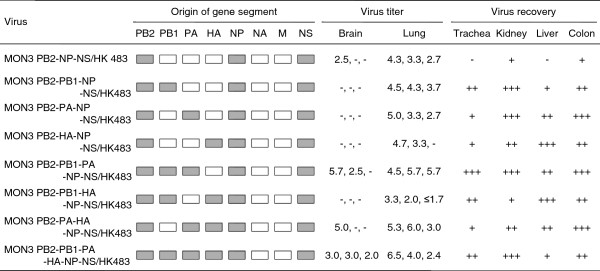
Method: To understand the mechanism whereby MON3 shows high pathogenicity in ducks, HK483, MON3, and twenty-four reassortants generated between these two H5N1 viruses were compared for their pathogenicity in domestic ducks.

Results: None of the ducks infected with MON3-based single-gene reassortants bearing the PB2, NP, or NS gene segment of HK483 died, and HK483-based single-gene reassortants bearing PB2, NP, or NS genes of MON3 were not pathogenic in ducks, suggesting that multiple gene segments contribute to the pathogenicity of MON3 in ducks. All the ducks infected with the reassortant bearing PB2, PA, HA, NP, and NS gene segments of MON3 died within five days post-inoculation, as did those infected with MON3. Each of the viruses was assessed for replication in ducks three days post-inoculation. MON3 and multi-gene reassortants pathogenic in ducks were recovered from all of the tissues examined and replicated with high titers in the brains and lungs.

Conclusion: The present results indicate that multigenic factors are responsible for efficient replication of MON3 in ducks. In particular, virus growth in the brain might correlate with neurological dysfunction and the disease severity.


Figures

Figure 1
Figure 1
Pathogenicity of HK483, MON3, and single-gene reassortants in 2-week-old ducks. HK483, MON3, and a series of reassortants between HK483 and MON3 were generated by reverse genetics. Each of the viruses was inoculated into 2-week-old ducks and clinical symptoms of challenged ducks were observed over 14 dpi. White and gray boxes indicate the derivation of virus gene segments from HK483 and MON3, respectively. *: number of dead birds/number of examined ducks. **: mean death dpi.
Figure 2
Figure 2
Pathogenicity of multi-gene reassortants in 2-week-old ducks. A series of multi-gene reassortants between HK483 and MON3 was generated by reverse genetics. Each of the viruses was inoculated into 2-week-old ducks and clinical symptoms of challenged ducks were observed over 14 dpi. White and gray boxes indicate the derivation of virus gene segments from HK483 and MON3, respectively. *: number of dead birds/number of examined ducks. **: mean death dpi.
Figure 3
Figure 3
Comparison of virus replication in 2-week-old ducks between HK483 and MON3. Three days post-inoculation, the brains, tracheas, lungs, kidneys, livers, and colons were collected. Viruses in each of the tissue samples were titrated in 10-day-old embryonated chicken eggs and titers were expressed as EID50/g of tissues. The mean virus titers in each of the tissues and standard deviations are shown. The lower limit of detection was 101.5 EID50/g. Virus titers were statistically analyzed by Student’s paired t-test. Significant differences are indicated by asterisks (** p < 0.001, * p < 0.05).
Figure 4
Figure 4
Replication of single-gene reassortants in 2-week-old ducks. A series of single-gene reassortants between HK483 and MON3 was generated by reverse genetics. Each of the viruses was inoculated into 2-week-old ducks and tissue samples were collected on 3 dpi. Suspension of each tissue sample was inoculated into 10-day-old embryonated eggs for virus titration or recovery. White and gray boxes indicate the derivation of virus gene segments from HK483 and MON3, respectively. Virus titers in tissues were expressed as log EID50/g and the lower limit of detection was 101.5 EID50/g. +, ++, and +++: viruses were recovered from one, two, and three of three birds tested, respectively. -: viruses were not detected from tissue samples.
Figure 5
Figure 5
Replication of multi-gene reassortants in 2-week-old ducks. A series of multi-gene reassortants between HK483 and MON3 was generated by reverse genetics. Each of the viruses was inoculated into 2-week-old ducks and tissue samples were collected on 3 dpi. Suspension of each tissue sample was inoculated into 10-day-old embryonated eggs for virus titration or recovery. White and gray boxes indicate the derivation of virus gene segments from HK483 and MON3, respectively. Virus titers in tissues were expressed as log EID50/g and the lower limit of detection was 101.5 EID50/g. +, ++, and +++: viruses were recovered from one, two, and three of three birds tested, respectively. -: viruses were not detected from tissue samples.

References

    1. Kawaoka Y, Cox NJ, Haller O, Hongo S, Kaverin N, Klenk HD, Lamb RA, McCauley J, Palese P, Rimstad E, Webster RG. In: Virus Taxonomy. 8. Fauquet CM, Mayo MA, Maniloff J, Desselberger U, Ball LA, editor. California: Elsevier Academic Press; 2005. Family Orthomyxoviridae; pp. 681–693.
    1. Fouchier RA, Munster V, Wallensten A, Bestebroer TM, Herfst S, Smith D, Rimmelzwaan GF, Olsen B, Osterhaus AD. Characterization of a novel influenza A virus hemagglutinin subtype (H16) obtained from black-headed gulls. J Virol. 2005;79:2814–2822. doi: 10.1128/JVI.79.5.2814-2822.2005. - DOI - PMC - PubMed
    1. Ito T, Okazaki K, Kawaoka Y, Takada A, Webster RG, Kida H. Perpetuation of influenza A viruses in Alaskan waterfowl reservoirs. Arch Virol. 1995;140:1163–1172. doi: 10.1007/BF01322743. - DOI - PubMed
    1. Okazaki K, Takada A, Ito T, Imai M, Takakuwa H, Hatta M, Ozaki H, Tanizaki T, Nagano T, Ninomiya A, Demenov VA, Tyaptirganov MM, Karatayeva TD, Yamnikova SS, Lvov DK, Kida H. Precursor genes of future pandemic influenza viruses are perpetuated in ducks nesting in Siberia. Arch Virol. 2000;145:885–893. doi: 10.1007/s007050050681. - DOI - PubMed
    1. Kida H, Yanagawa R, Matsuoka Y. Duck influenza lacking evidence of disease signs and immune response. Infect Immun. 1980;30:547–553. - PMC - PubMed

Publication types

MeSH terms

Substances