. 2013 Feb 1:11:12.
doi: 10.1186/1741-7007-11-12.

MST1, a key player, in enhancing fast skeletal muscle atrophy

Affiliations

MST1, a key player, in enhancing fast skeletal muscle atrophy

Bin Wei et al. BMC Biol. .

Abstract

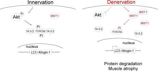
Background: Skeletal muscle undergoes rapid atrophy upon denervation and the underlying mechanisms are complicated. FOXO3a has been implicated as a major mediator of muscle atrophy, but how its subcellular location and activity is controlled during the pathogenesis of muscle atrophy remains largely unknown. MST1 (Mammalian Sterile 20-like kinase 1) is identified as a central component of the Hippo signaling pathway. MST1 has been shown to mediate phosphorylation of FOXO3a at Ser207. Whether this MST1-FOXO signaling cascade exerts any functional consequence on cellular homeostasis remains to be investigated.

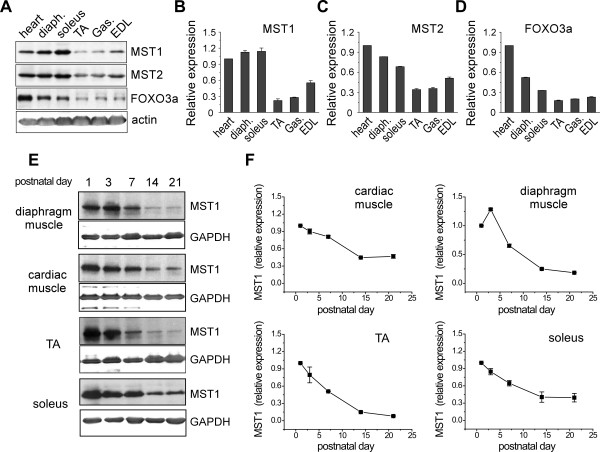
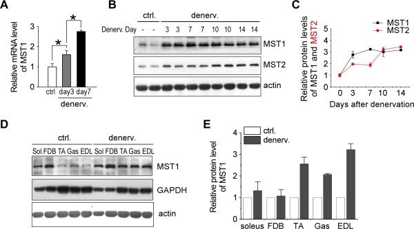
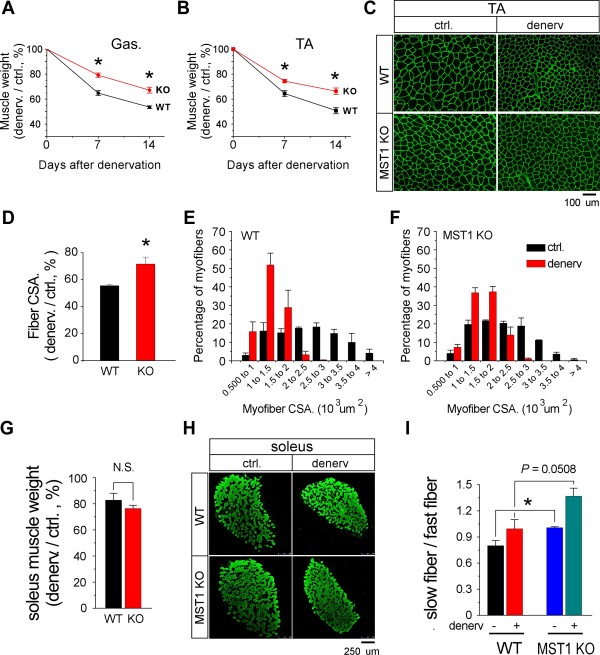
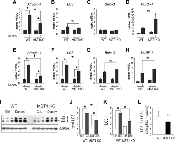
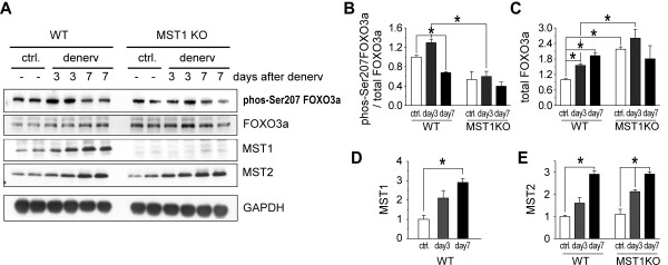
Result: We identified that MST1 kinase was expressed widely in skeletal muscles and was dramatically up-regulated in fast- but not slow-dominant skeletal muscles immediately following denervation. The results of our histological and biochemical studies demonstrated that deletion of MST1 significantly attenuated denervation-induced skeletal muscle wasting and decreased expression of Atrogin-1 and LC3 genes in fast-dominant skeletal muscles from three- to five-month-old adult mice. Further studies indicated that MST1, but not MST2, remarkably increased FOXO3a phosphorylation level at Ser207 and promoted its nuclear translocation in atrophic fast-dominant muscles.

Conclusions: We have established that MST1 kinase plays an important role in regulating denervation-induced skeletal muscle atrophy. During the early stage of muscle atrophy, the up-regulated MST1 kinase promoted progression of neurogenic atrophy in fast-dominant skeletal muscles through activation of FOXO3a transcription factors.


Figures

Figure 1
Figure 1
MST1 is expressed in striated muscles and enriched at early stage of postnatal muscle development. A. Expressions of MST1/2 and FOXO3a in striated muscles from three-month-old adult wild type mice were identified by Western blotting. B-D. Densitometry analysis showed the expressions of MST1/2 kinases were enriched in cardiac muscle, diaphragm as well as soleus muscles, which contain significant amount of type I fibers. E. Representative blotting shows protein level of MST1 was decreased during postnatal development in diaphragm, cardiac, TA and soleus muscles. F. Quantification of muscular MST1 expression during postnatal development.
Figure 2
Figure 2
MST1 kinase is dramatically increased during denervation-induced atrophy in fast-dominant muscles from adult mice. A. Quantitative PCR showed mRNA of MST1 was continuously increased three and seven days after denervation. B and C. Western blotting and densitometry showed MST1 and MST2 protein levels were both increased during the process of denervation-induced atrophy in TA muscle. D and E. Seven days after denervation, MST1 protein levels were dramatically increased in multiple fast-dominant muscles (TA, Gas and EDL), but not in slow-dominant soleus and FDB muscles. *P < 0.05 by two-tailed Student's test.
Figure 3
Figure 3
MST1 deficiency protects fast- but not slow-dominant skeletal muscles against denervation-induced atrophy. Sciatic denervation was performed on three- to five-month-old adult WT and MST1 KO mice littermates. Three pairs of WT and MST1 KO mice were sacrificed at Day 7 after denervation and the other three pairs were examined at Dday 14. A. Mass of denervated gastrocnemius muscles from MST1 deficient mice remained at 79.18% ± 1.73% (on Day 7) and 67.14% ± 2.37% (on Day 14) of the contralateral muscles, both of which were significantly higher than 64.83% ± 2.01% and 53.53% ± 1.09% in WT mice, respectively. B. Seven and 14 days after denervation, mass of denervated TA muscles from MST1 KO mice remained at 74.43% ± 1.36% and 66.35% ± 2.56% of contralateral levels, whereas WT TA muscle mass remained at 64.47% ± 2.4% and 50.81% ± 2.5%, respectively. C. Representative images of α-laminin staining on contralateral and denervated TA muscle cryosections, indicating myofiber size was better preserved in MST1 deficient TA muscles at Day 14 after denervation. D. Fourteen days after denervation, averaged myofiber cross-sectional area of denervated TA muscle decreased to 71.61% ± 5.11% of the contralateral level in MST1 KO mice and 55.56% ± 0.87% in WT mice. E and F. TA myofiber size distribution showed a suppressed leftward shift in MST1 deficient TA muscles (F) 14 days after denervation, indicating less atrophy compared with WT muscles (E). G. Fourteen days after denervation, WT soleus muscle mass remained at 82.78% ± 5.32% of contralateral and MST1 deficient soleus muscle mass remained at 76.43% ± 2.45% of its contralateral level. H. Representative images of MHC I staining on contralateral and post-denervated Day 14 soleus muscles from WT and MST1 KO mice. WT and MST1 deficient soleus muscles developed similar level of atrophy 14 days after denervation. I. The ratio of slow-to-fast fiber content was significantly higher in MST1 deficient contralateral soleus muscles than that of WT muscles. 14 days after denervation, the ratios of slow-to-fast fiber content in WT and MST1 deficient soleus muscle were both increased. * P < 0.05 compared with wild type by two-tailed Student's test.
Figure 4
Figure 4
Expression of atrophy related genes in denervated gastrocnemius muscles from WT and MST1 KO mice. A-D. Quantitative PCR results showed three days after denervation, Atrogin-1 mRNA levels in WT and MST1 deficient gastrocnemius muscle were both increased (by 4.57 ± 0.70 folds and 2.68 ± 0.28 folds of the contralateral level, respectively). The increases in mRNA levels of LC-3 (1.76 ± 0.35 and 1.54 ± 0.15, WT vs. MST1 KO) and Bnip-3 were both less than two-fold and there is no significant difference in the WT and MST1 deficient muscles. MuRF-1 mRNA levels were upregulated to 9.51 ± 0.88 and 24.07 ± 7.68 folds in WT and MST1 deficient muscles, respectively. E-H. At Day 7 after denervation, the mRNA levels of muscle atrophy-related genes were significantly increased in both WT and MST1 deficient denervated muscles. The mRNA levels of Atrogin-1 and LC3 in denervated WT muscles (4.45 ± 0.47 and 4.43 ± 0.26 folds increased, respectively) were both significantly higher than that of MST1 KO mice (1.82 ± 0.12 folds and 2.91 ± 0.06 folds, respectively). Bnip-3 and MuRF-1 mRNA levels were also increased in denervated WT and MST1 deficient muscles but showed no differences between the genotypes. I-L. The protein levels of total LC3 and lapidated form (LC3 II) were both increased in MST1 deficient and WT muscles. Total LC3 protein was increased at 3.95 ± 0.37 folds in WT denervated muscle and 2.65 ± 0.48 folds in MST1 deficient denervated muscles. The LC3 II protein level in WT denervated muscle was increased 3.95 ± 0.31 folds and 2.44 ± 0.54 folds in MST1 deficient muscles, respectively. The ratio of LC3 II/LC3 I was 1.68 ± 0.20 and 1.31 ± 0.12 in WT and MST1 deficient muscles after denervation. * P < 0.05 by two-tailed Student's test.
Figure 5
Figure 5
MST1 phosphorylates FOXO3a at Ser207 during muscle atrophy. A. Representative western blotting of MST1/2 and FOXO3a in atrophic TA muscles. B. Phosphorylation of FOXO3a at serine 207 by MST1 kinase was increased (by 29.5% ± 7.40%) in WT TA muscles but not in MST1 deficient muscles at denervation Day 3. FOXO3a Ser207 phosphorylation was decreased both in WT and MST1 KO muscles at Day 7 after denervation. C. Total FOXO3a protein level was continuously increased at Day 3 and Day 7 after denervation in WT TA muscles. D and E. Both the expression levels of MST1 and MST2 kinases were increased in atrophic muscles. MST2 protein levels were similarly increased in wild type and MST1 deficient muscles and WT muscles after denervation. At Dday 7 after denervation, comparing with the contralateral, the protein levels of MST2 kinase were increased 2.91 ± 0.15 and 2.92 ± 0.10 folds in the atrophic TA muscles obtained from WT and MST1 KO mice, respectively. * P < 0.05 by two-tailed Student's test.
Figure 6
Figure 6
MST1 promotes nuclear translocation of FOXO3a through phosphorylation at Ser207. A and C. Representative immunostaining using pan-FOXO3a and Ser207-phosphorylated FOXO3a antibodies on TA muscle sections obtained on denervation Day 3. B. At Day 3 after denervation, 41.46% ± 3.07% and 27.51% ± 2.28% of the total myofiber nucleus were stained with FOXO3a in WT and MST1 deficient TA muscles, respectively. D. At Day 3 after denervation, 32.63% ± 1.82% and 12.10% ± 1.49% of total TA muscle nuclei stained with serine 207-posphorylated FoxO3a in WT and MST1 mice, respectively. *** P < 0.0001 by two-tailed Student's test..
Figure 7
Figure 7
Schematic diagram of MST1's function in mediating fast skeletal muscle atrophy. Within an innervated myofiber, Akt kinase is active and MST1 kinase amount is restricted at low level. Thus FOXO3a is phosphorylated by Akt at multiple sites (Thr32, Ser253 and Ser315) [53], and is restricted within the myofiber cytosol through interaction with 14-3-3 proteins. Denervation induces rapid up-regulation of MST1 kinase. MST1 phosphorylates FOXO3a at Ser207 and causes its translocation into myofiber nucleus to activate expression of LC3 and Atrogin-1 genes, which leads to rapid muscle mass loss during the progression of muscle atrophy.

Similar articles

Cited by

References

    1. Tidball JG. Mechanical signal transduction in skeletal muscle growth and adaptation. J Appl Physiol. 2005;98:1900–1908. doi: 10.1152/japplphysiol.01178.2004. - DOI - PubMed
    1. Andersson DC, Betzenhauser MJ, Reiken S, Meli AC, Umanskaya A, Xie W, Shiomi T, Zalk R, Lacampagne A, Marks AR. Ryanodine receptor oxidation causes intracellular calcium leak and muscle weakness in aging. Cell Metab. 2011;14:196–207. doi: 10.1016/j.cmet.2011.05.014. - DOI - PMC - PubMed
    1. Mourtzakis M, Bedbrook M. Muscle atrophy in cancer: a role for nutrition and exercise. Appl Physiol Nutr Metab. 2009;34:950–956. doi: 10.1139/H09-075. - DOI - PubMed
    1. Penna F, Bonetto A, Muscaritoli M, Costamagna D, Minero VG, Bonelli G, Rossi Fanelli F, Baccino FM, Costelli P. Muscle atrophy in experimental cancer cachexia: is the IGF-1 signaling pathway involved? Int J Cancer. 2010;127:1706–1717. doi: 10.1002/ijc.25146. - DOI - PubMed
    1. Carvalho RF, Castan EP, Coelho CA, Lopes FS, Almeida FL, Michelin A, de Souza RW, Araujo JP Jr, Cicogna AC, Dal Pai-Silva M. Heart failure increases atrogin-1 and MuRF1 gene expression in skeletal muscle with fiber type-specific atrophy. J Mol Histol. 2010;41:81–87. doi: 10.1007/s10735-010-9262-x. - DOI - PubMed

Publication types

MeSH terms

LinkOut - more resources