. 2013 Feb 4:14:9.
doi: 10.1186/2050-6511-14-9.

Effects of early life exposure to ultraviolet C radiation on mitochondrial DNA content, transcription, ATP production, and oxygen consumption in developing Caenorhabditis elegans

Affiliations

Effects of early life exposure to ultraviolet C radiation on mitochondrial DNA content, transcription, ATP production, and oxygen consumption in developing Caenorhabditis elegans

Maxwell C K Leung et al. BMC Pharmacol Toxicol. .

Abstract

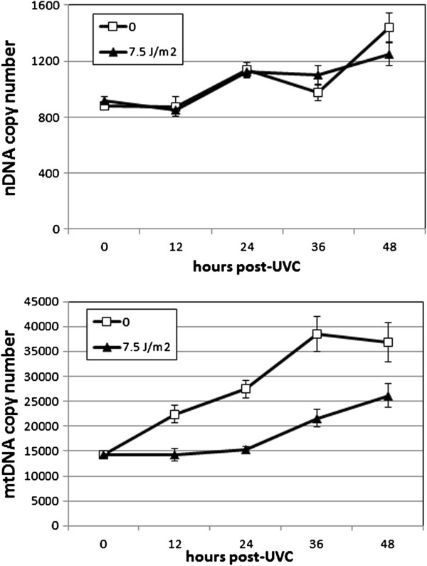
Background: Mitochondrial DNA (mtDNA) is present in multiple copies per cell and undergoes dramatic amplification during development. The impacts of mtDNA damage incurred early in development are not well understood, especially in the case of types of mtDNA damage that are irreparable, such as ultraviolet C radiation (UVC)-induced photodimers.

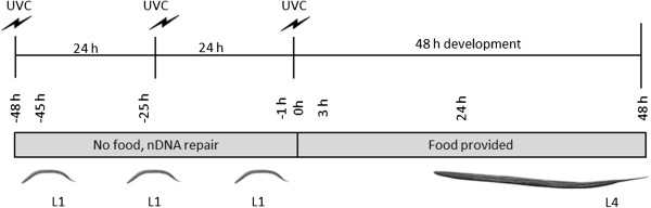
Methods: We exposed first larval stage nematodes to UVC using a protocol that results in accumulated mtDNA damage but permits nuclear DNA (nDNA) repair. We then measured the transcriptional response, as well as oxygen consumption, ATP levels, and mtDNA copy number through adulthood.

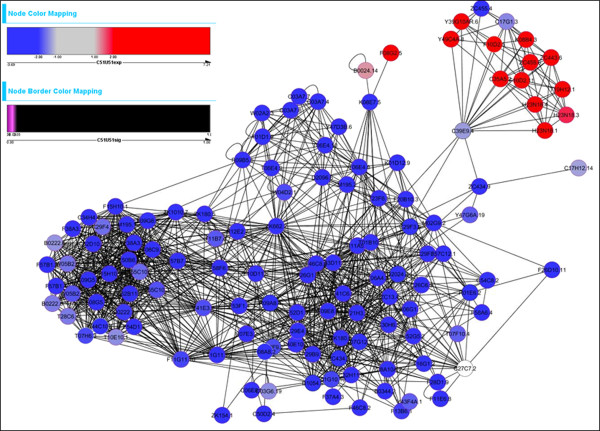
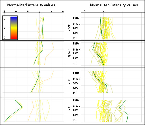
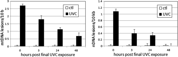
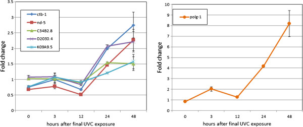
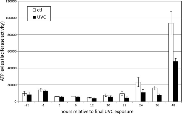
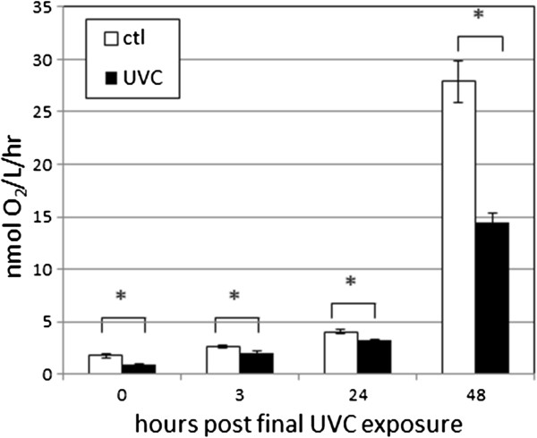
Results: Although the mtDNA damage persisted to the fourth larval stage, we observed only a relatively minor ~40% decrease in mtDNA copy number. Transcriptomic analysis suggested an inhibition of aerobic metabolism and developmental processes; mRNA levels for mtDNA-encoded genes were reduced ~50% at 3 hours post-treatment, but recovered and, in some cases, were upregulated at 24 and 48 hours post-exposure. The mtDNA polymerase γ was also induced ~8-fold at 48 hours post-exposure. Moreover, ATP levels and oxygen consumption were reduced in response to UVC exposure, with marked reductions of ~50% at the later larval stages.

Conclusions: These results support the hypothesis that early life exposure to mitochondrial genotoxicants could result in mitochondrial dysfunction at later stages of life, thereby highlighting the potential health hazards of time-delayed effects of these genotoxicants in the environment.


Figures

Figure 1
Figure 1
Experimental design. Liquid-hatched L1 stage C. elegans were exposed to 7.5 J/m2 UVC over 48 h, in the absence of food, permitting nDNA repair but accumulation of mtDNA damage [15]. Nematodes were then placed on food plates and followed for another 48 h. We measured mRNA levels, genome copy number, DNA damage, ATP levels, and oxygen consumption at multiple times during and after the UVC exposures. All times are given relative to the final dose and transferral to food (= “0 h”). For example, mRNA sampled immediately prior to the second dose of UVC would be described as sampled at “-25 h”, and “3 h” if it were sampled 3 h after the final UVC exposure. Representative examples are presented to orient the reader.
Figure 2
Figure 2
At 3 h, the combination UVC + EtBr treatment resulted in dramatic changes in expression of networked genes. The blue genes are downregulated after the combination treatment and comprise genes belonging to body morphogenesis and other developmental processes. Most red genes (upper right cluster) are part of lipid and carbohydrate metabolic processes.
Figure 3
Figure 3
EtBr and UVC exposures resulted in decreased expression of many oxidative phosphorylation genes (top panel; sucg-1 highlighted in green), but increased expression of some glyoxylate genes (bottom panel; icl-1 highlighted in green). Red-blue scale coloration is based on comparison to mRNA levels in control samples at −45 h. Normalized intensity values are on a binary log scale (i.e. “1” indicates a 2-fold change, “2” a 4-fold change, etc.). n = 4–6.
Figure 4
Figure 4
Exposure to UVC resulted in persistent mtDNA damage in larval C. elegans. Main effects of treatment, time, genome, and all interactions were significant (p ≤ 0.0002). At 0, 3, and 24 h after the last exposure, DNA damage was statistically significant in both genomes. At 48 h, DNA damage was detected in mtDNA (p = 0.0009) but not nDNA (p = 0.82). Note different y-axis scales. n = 4–11 in two experiments.
Figure 5
Figure 5
Exposure to UVC did not cause a detectable change in nDNA copy number, but decreased mtDNA copy number at later time points. Effect on nDNA: p = 0.82 for main effect of UVC, p = 0.37 for interaction of UVC with time, p < 0.0001 main effect of time. Effect on mtDNA: p < 0.0001 for main effects of UVC and time, p = 0.15 for interaction. n = 8–20 (3 or 4 separate experiments).
Figure 6
Figure 6
Exposure to UVC reduced ctb-1 and nd-5 expression at early timepoints, but resulted in higher levels of all mRNAs at later times (wild-type N2 strain).ctb-1 and nd-5 are mtDNA-encoded, and C34B2.8, D2030.4, K09A9.5, polg-1 are nDNA-encoded, mitochondrial proteins. polg-1 is graphed separately to avoid obscuring the smaller fold-changes observed in the other genes; note different y-axes. p < 0.0001 for all main and interactive effects, 3 factor ANOVA. Fold change is relative to the mRNA of the same gene at the same time without UVC exposure. n = 3–6.
Figure 7
Figure 7
Exposure to UVC reduced ATP levels at later larval stages in developing C. elegans. The main effects of UVC and time were significant (p = 0.003 and p < 0.0001 respectively), but their interaction was not, precluding comparisons at specific timepoints. n = 5–7 separate experiments; 5 separate measurements per experiment were pooled for each “n”.
Figure 8
Figure 8
Exposure to UVC reduced oxygen consumption in larval C. elegans. n = 4–6 in 3 experiments. The effects of time and UVC and their interactions were all significant at p < 0.0001 (ANOVA); all FPLSD comparisons for the effect of UVC at individual timepoints were significant at p ≤ 0.001 (indicated by asterisks).

References

    1. Schmidt CW. Mito-conundrum unraveling environmental effects on mitochondria. Environ Heal Perspect. 2010;118:A292–A297. doi: 10.1289/ehp.118-a292. - DOI
    1. Shaughnessy DT, Worth L, Lawler CP, McAllister KA, Longley MJ, Copeland WC. Meeting report: Identification of biomarkers for early detection of mitochondrial dysfunction. Mitochondrion. 2010;10:579–581. doi: 10.1016/j.mito.2010.02.001. - DOI - PubMed
    1. Larsen NB, Rasmussen M, Rasmussen LJ. Nuclear and mitochondrial DNA repair: similar pathways? Mitochondrion. 2005;5:89–108. doi: 10.1016/j.mito.2005.02.002. - DOI - PubMed
    1. Cohen BH. Pharmacologic effects on mitochondrial function. Dev Disabil Res Rev. 2010;16:189–199. doi: 10.1002/ddrr.106. - DOI - PubMed
    1. Gomez C, Bandez MJ, Navarro A. Pesticides and impairment of mitochondrial function in relation with the parkinsonian syndrome. Front Biosci. 2007;12:1079–1093. doi: 10.2741/2128. - DOI - PubMed

Publication types

MeSH terms

LinkOut - more resources