Review
. 2013 Mar;21(3):145-56.
doi: 10.1016/j.tim.2012.12.004. Epub 2013 Jan 30.

Eukaryotic virulence determinants utilize phosphoinositides at the ER and host cell surface

Affiliations
Review

Eukaryotic virulence determinants utilize phosphoinositides at the ER and host cell surface

Rays H Y Jiang et al. Trends Microbiol. 2013 Mar.

Abstract

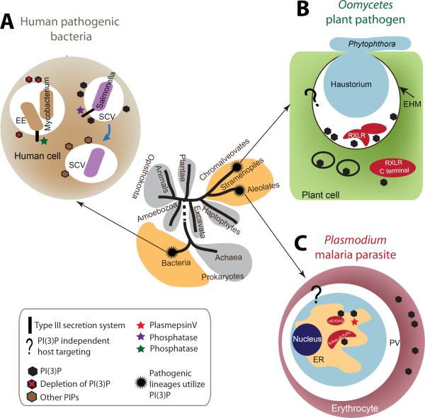
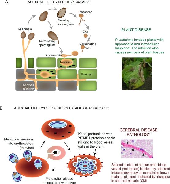
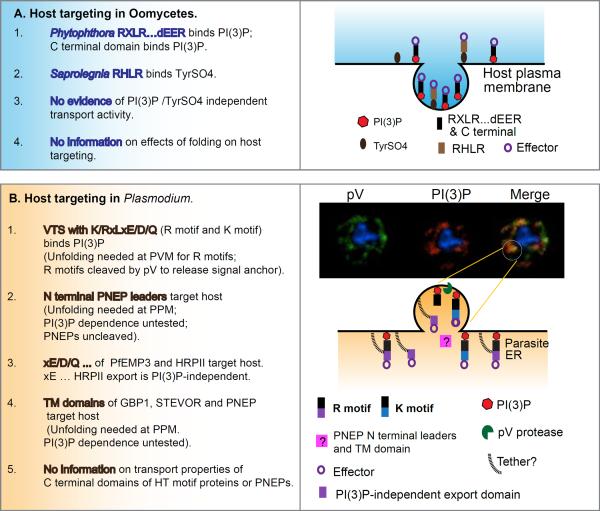
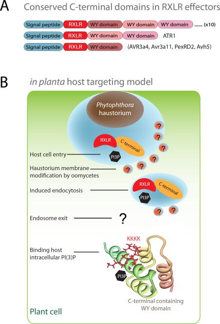
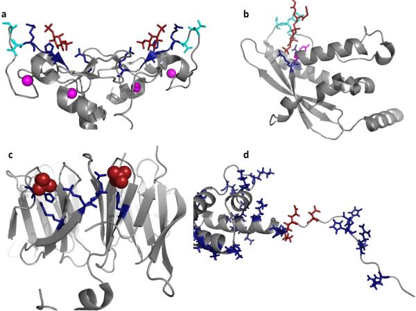
Similar to bacteria, eukaryotic pathogens may utilize common strategies of pathogenic secretion, because effector proteins from the oomycete Phytophthora infestans and virulence determinants from the human malaria parasite Plasmodium falciparum share a functionally equivalent host-cell-targeting motif (RxLR-dEER in P. infestans and RxLxE/D/Q in P. falciparum). Here we summarize recent studies that reveal that the malarial motif may function differently than previously envisioned. Binding of the lipid phosphatidylinositol 3-phosphate [PI(3)P] is a critical step in accessing the host for both pathogens, but occurs in different locations. Nanomolar affinity for PI(3)P by these short amino acid motifs suggests that a newly identified mechanism of phosphoinositide binding that unexpectedly occurs in secretory locations has been exploited for virulence by diverse eukaryotic pathogens.


Figures

Figure 1
Figure 1. Phylogenetic position, modulation of PI(3)P and host targeting in different intracellular pathogens
The tree of major life domains shown is based on the analysis of [55]. Pathogenic lineages that utilize PI(3)P for host targeting and host modulation are chromalveolates, alveolates and bacteria. The life domains to which they belong are colored yellow. Host targeting refers to the process by which intracellular pathogens deliver pathogen-produced enzymes, toxins and other proteins directly into host cytoplasm and membrane. Bacterial pathogens use a type III secretion system to inject effectors into host cells. Eukaryotic pathogens utilize endocytic pathways to deliver proteins. (a) Intracellular bacteria use type III effectors to modulate PI(3)P levels for survival and replication within vacuoles inside the host cell cytoplasm. Mycobacterium tuberculosis decreases PI(3)P levels to arrest endosome maturation, avoiding destruction by the host. Salmonella transiently increases PI(3)P levels to facilitate invasion and biogenesis of the vacuoles for intracellular survival. (b) Plant pathogenic oomycetes partly grow into plant cells with a structure called the haustorium. Host targeting utilizes PI(3)P at the extra haustorial membrane surrounding the haustorium to enable RxLR-dEER effector entry. How effectors are delivered across the extra haustorial membrane into the host cell is not known (and thus shown by a question mark), but once in the cytoplasm, their C-terminal domain(s) can bind cytoplasmic PI(3)P to modulate host physiology. (c) The malaria parasite Plasmodium survives in a membranous structure in erythrocytes. Host targeting starts at the parasite ER with binding of PI(3)P by a host targeting motif R/KxLxE/D/Q. Plasmepsin V is an aspartic protease that does not cleave KxL motifs. However it is detected in association with PI(3)P (see Figure 3) cleaves after the RxL to yield xE/D/Q, which does not bind PI(3)P but supports export by unknown mechanisms (depicted by question mark). In oomycetes, PI(3)P-independent host targeting pathways also exist. Vaid et al. [56] report the presence of PI(3)P in the infected erythrocyte. Abbreviations: EE, early endosome; SCV, Salmonella-containing vacuole; EHM, extra haustorial membrane; ER, endoplasmic reticulum; PV, parasitophorous vacuole.
Figure 2
Figure 2. Life cycles and pathologies of Phytophthora and Plasmodium
(a) P. infestans is able to initiate an infection with both sporangia and zoospores. The pathogen forms an appressorium and penetrates plant leaf cells. Subsequently, a haustorium is developed within host cytoplasm for nutrient acquisition and host immunity modulation. Effectors (black bricks) are trafficked into host cells from the haustoria. Later during infection, P. infestans causes necrosis in plant tissue and sporulates to form new sporangia. The photo of infected potato leaves with sporulating P. infestans is kindly provided by Klaas Bouwmeester and Francine Govers. (b) Asexual, P. falciparum blood stage infection begins when the extracellular merozoite invades the erythrocyte, a process which although complex, is completed in minutes. Effectors (black bricks) are exported across the vacuolar membrane that surrounds the intracellular parasite. Subsequent parasite development, maturation and emergence of merozoites takes ~48 h. In the second half of the asexual cycle, exported parasite effectors assemble in electron dense `knobs' that display the cell surface parasite protein PfEMP1. PfEMP1s are endothelial adhesins that enable infected erythrocytes to bind to the walls of blood vessels. In the brain this can lead to inflammation as well as occlusion of blood vessels (as seen in the adjacent tissue section) and the fatal, neurological pathology of cerebral malaria. The photo is kindly provided by Dan Millner.
Figure 3
Figure 3. Summary of transport properties of (a) oomycete and (b) Plasmodium protein domains
(a) In Phytophthora, leaders containing RxLR- dEER and/or C terminal domains (black bars) of effector proteins (purple circles) binds the lipid PI(3)P (red dots) at the host plasma membrane resulting in their endosomal internalization. In Saprolegnia, leaders containing RHLR (brown bars) binds tyrosine sulfate (TyrSO4; grey dot, a post translational modification of host proteins) to also undergo endosomal internalization. Presently, there is no evidence of host targeting properties independent of binding PI(3)P or TyrSO4 or protein folding, such as seen in Plasmodium. (b) In Plasmodium, leaders known as vacuolar translocation sequences (VTS) containing a host targeting motif KxLxE/D/Q (black/blue bar) or RxLxE/D/Q (black/purple bar) bind PI(3)P (red dots) in the ER lumen. Micrographs in the right hand side top panel show distribution of the lipid PI(3)P in the ER lumen (red) and a parasite protease plasmepsin V (pV; green) colocalized (yellow/orange) in membrane domains in the ER. This suggests a model (shown immediately below) for a PI(3)P enriched vesicle (containing pV) emerging from the ER. In this regard, PI(3)P-dependent exit from the ER is topologically equivalent to PI(3)P-dependent endosomal uptake at the plasma membrane. Proteins with RxLxE/D/Q may be cleaved by the protease pV in the emerging PI(3)P vesicle, (which is reinforced by the finding that the specificity of PI(3)P binding mimics that of pV mediated cleavage). The xE/D/Q fragments of PfEMP3 and PfHRPII (purple rectangle) also have intrinsic export activity suggesting a second mechanism of inclusion and retention of soluble proteins (indicated by black tethers). N terminal leaders lacking K/RxL domains called PNEPs (pink rectangles) appear to share a common export mechanism whose dependence on PI(3)P has not been tested (and thus indicated by a question mark). Unlike Phytophthora, C-terminal domains of plasmodial effectors have not yet been shown to have intrinsic PI(3)P binding or export activity and parasite host targeting is sensitive to protein folding.
Figure 4
Figure 4. Conserved RxLR-dEER effector structure fold and different sites for PI(3)P binding
(a) RxLR effector domain architecture. RxLR effectors have an N-terminal secretory leader, a host targeting RxLR motif and conserved C-terminal domains that are often arranged into tandem repeats. (b) Host targeting model and involvement of PI(3)P at different cellular sites. The N-terminal RxLR motifs play an important role in host cell entry at host plasma membrane; and in vitro recombinant proteins of Avr1b bind PI(3)P. The C-terminal region has a conserved effector fold and is able to suppress host defense responses. The positive patches in the C-terminal domain of Avr1b and Avr3a are important for PI(3)P and PI(4)P binding. The Avr1b C-terminal structure is used in the illustration. Several important conceptual steps such as modification of membrane by pathogen are postulated and indicated with question marks.
Figure 5
Figure 5. Mechanisms and models of PI(3)P binding by FYVE, PX, PROPPIN, RxLR and WY domains
PI(3)P binding has been attributed to at least three distinct eukaryotic domains including FYVE, PX, and PROPPINS while pathogens utilize the RxLR or RxLxE/D/Q motif and/or WY domains. (a) The homodimeric structure of the EEA1 FYVE domain is shown (PDB ID: 1JOC), which binds PI(3)P (shown in red) in a sideways fashion where both electrostatic and H-bonding with PI(3)P can be optimized while achieving hydrophobic interactions through the turret loops [39]. Cationic residues that coordinate the PI(3)P headgroup are shown in blue, hydrophobic residues that penetrate into the hydrocarbon portion of the membrane are highlighted in cyan, and the zinc ions coordinated by the FYVE domains for structural stability are shown in magenta. (b) The p40phox PX domain (PDB ID: 1H6H) X-ray structure was solved bound to PI(3)P. Here, two critical arginine residues (shown in blue) mediate the recognition of PI(3)P, while a tyrosine residue (magenta) forms the bottom of the PI(3)P binding pocket. The PI(3)P binding pocket is deeper than that observed for the FYVE domain and harbors two loop regions that have been shown to facilitate nonspecific electrostatic interactions and membrane penetration (hydrophobic residues in cyan) [57]. (c) The yeast PROPPIN structure from Hsv2 (Homologous with SVP1), which is a key autophagic sensor protein, was solved bound to two sulfate ions (PDB ID: 4EXV). PROPPINs are PI(3)P sensors necessary for autophagy and are conserved from yeast to humans. The PROPPIN structure is a β-propeller composed of seven blades where two PI(3)P binding sites (sulfate ions in red and coordinating residues in blue) were discovered on blades five and six. In a similar manner to the FYVE and PX domains, a hydrophobic loop between blades five and six promotes membrane penetration of Hsv2, which is essential to act in concert with PI(3)P binding for membrane recognition and full autophagic function in yeast. (d) Shown is the N-terminal unstructured region and WY domain of Avr1b from P. sojae, which was modeled and generated using the available P. infestans Avr3a structure (PDB ID: 2LC2) [25]. The N-terminus harbors the RxLR motif (Arg residues in blue on the left) as well as number of other cationic (blue) and anionic (red) residues that may be critical tophosphoinositide binding. The WY domain of Avr1b is shown to highlight the cationic nature of this module (Arg, Lys and His shown in blue). In contrast to FYVE, PX, and PROPPINs a distinct phosphoinositide binding pocket or region is not obvious and nonspecific electrostatic association may occur based upon lipid charge.

Similar articles

Cited by

References

    1. Di Paolo G, De Camilli P. Phosphoinositides in cell regulation and membrane dynamics. Nature. 2006;443:651–657. - PubMed
    1. Behnia R, Munro S. Organelle identity and the signposts for membrane traffic. Nature. 2005;438:597–604. - PubMed
    1. Dai S, et al. Bacteria-generated PtdIns(3)P recruits VAMP8 to facilitate phagocytosis. Traffic. 2007;8:1365–1374. - PubMed
    1. Galan JE. Salmonella interactions with host cells: Type III secretion at work. Ann. Rev. Cell Dev. Biol. 2001;17:53–86. - PubMed
    1. Mallo GV, et al. SopB promotes phosphatidylinositol 3-phosphate formation on Salmonella vacuoles by recruiting Rab5 and Vps34. J Cell Biol. 2008;182:741–752. - PMC - PubMed

Publication types