Review
. 2013 Jun;23(3):294-303.
doi: 10.1016/j.conb.2013.01.017. Epub 2013 Jan 31.

Neuroscience of affect: brain mechanisms of pleasure and displeasure

Affiliations
Review

Neuroscience of affect: brain mechanisms of pleasure and displeasure

Kent C Berridge et al. Curr Opin Neurobiol. 2013 Jun.

Abstract

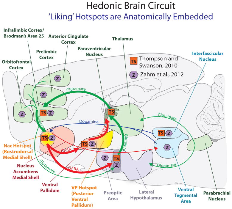
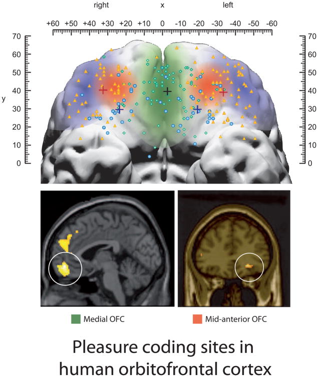
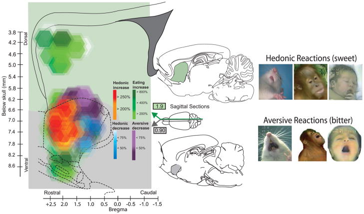
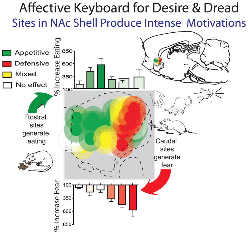
Affective neuroscience aims to understand how affect (pleasure or displeasure) is created by brains. Progress is aided by recognizing that affect has both objective and subjective features. Those dual aspects reflect that affective reactions are generated by neural mechanisms, selected in evolution based on their real (objective) consequences for genetic fitness. We review evidence for neural representation of pleasure in the brain (gained largely from neuroimaging studies), and evidence for the causal generation of pleasure (gained largely from brain manipulation studies). We suggest that representation and causation may actually reflect somewhat separable neuropsychological functions. Representation reaches an apex in limbic regions of prefrontal cortex, especially orbitofrontal cortex, influencing decisions and affective regulation. Causation of core pleasure or 'liking' reactions is much more subcortically weighted, and sometimes surprisingly localized. Pleasure 'liking' is especially generated by restricted hedonic hotspot circuits in nucleus accumbens (NAc) and ventral pallidum. Another example of localized valence generation, beyond hedonic hotspots, is an affective keyboard mechanism in NAc for releasing intense motivations such as either positively valenced desire and/or negatively valenced dread.


Figures

Figure 1
Figure 1
Hedonic hotspots and anatomical circuits that distinguish the nucleus accumbens hotspot in rostrodorsal medial shell as a unique site (anatomy based on Thompson and Swanson (TS symbol in orange boxes; 2010) and on Zahm and colleagues (Z symbol in purple hexagons; 2012). Thompson and Swanson [66] reported that the nucleus accumbens hotspot of rostrodorsal medial shell is uniquely embedded in its own closed-circuit corticolimbicpallidal-thalamocortical loop, connecting discrete input subregions and output subregions, and segregated from other parallel loops passing through other regions of medial shell. Zahm and colleagues suggested additional unique connections for the rostrodorsal hotspot [65]. GABAergic projections are indicated in red, hedonic hotspots are marked in yellow, glutamatergic projections are green, and dopaminergic projections are marked in blue. Figure by Daniel Castro, modified from [81].
Figure 2
Figure 2
Subjective pleasure is faithfully coded by orbitofrontal cortex (OFC) activations in people. Sensory pleasures appear most faithfully represented especially by a mid-anterior OFC site (orange). Pleasant sensations are also coded by activation in a medial strip of OFC (green), but the medial strip may not as faithfully track changes in pleasure as the orange mid-anterior site [37]. Smaller symbols show results of a large meta-analysis of 267 orbital areas, which indicatedthat a medial subregion of orbitofrontal cortex monitored learning and memory of reward values (green area and round blue dots), whereas a lateral orbitofrontal subregion monitored punishers (purple and orange triangles) [82]. Independently, posterior subregions of OFC represented complex or abstract reinforcers (such as money) whereas anterior subregions represented sensory rewards such as taste [82].
Figure 3
Figure 3
Detail of hedonic hotspot in nucleus accumbens for pleasure generation (sagittal view of medial shell and of neostriatum). This is a causation map: colors reflect hedonic or motivation consequences (on liking reactions or on food intake) of mu opioid agonist microinjections at each site (based originally on Peciña & Berridge, 2005). Red/orange symbols in the rostrodorsal hotspot show sites that caused doubling or higher levels of hedonic liking reactions to sucrose taste. By comparison, at caudal sites the same opioid microinjections only suppressed aversive disgust reactions to bitter quinine (purple; e.g., suppressed gapes), or bivalently suppressed both liking and disgust reactions (blue). Green sites denote increases in motivation wanting to eat without any hedonic change in either liking or disgust (enhanced motivation also extended through all red/purple/blue sites in nucleus accumbens). Modified from [81], based on data from [51].
Figure 4
Figure 4
Affective keyboard pattern in nucleus accumbens for releasing intense desire and/or dread. The keyboard pattern of intense motivated behaviors is revealed in the consequences of drug microinjections at various rostrocaudal sites in medial shell. Microinjections of drugs that relatively inhibit accumbens neurons via amino acid neurotransmitters (e.g., a GABA agonist or a glutamate antagonist) in turn may disinhibit or release motivation generating circuits in downstream target structures. Rostral green sites released stimulation of eating by up to 600% (desire only). Caudal red sites released purely increased fearful reactions at levels up to 600% over normal (dread only; escape attempts, distress calls, defensive bite attempts; spontaneous anti-predator treading/burying). Yellow sites released both desire and dread in the same rats during the same 1-hr test. Just as a keyboard has many notes, bars reflect the many graded mixtures of affective desire-dread released as microinjection sites move rostrocaudal location in medial shell (appetitive desire to eat at top; fearful dread reactions at bottom). Modified from [81], based on data from [77,78,80].

References

    1. Lindquist KA, Wager TD, Kober H, Bliss-Moreau E, Barrett LF. The brain basis of emotion: A meta-analytic review. The Behavioral and brain sciences. 2012;35:121–143. A thoughtful review of neuroimaging studies, concluding that specific emotions do not have different discrete neural substrates. - PMC - PubMed
    1. Leknes S, Tracey I. A common neurobiology for pain and pleasure. Nat Rev Neurosci. 2008;9:314–320. Penetrating review of brain mechanisms involved in both pleassures and displeasures. - PubMed
    1. Berridge KC, Kringelbach ML. Building a neuroscience of pleasure and well-being. Psychol Well Being. 2011;1:1–3. - PMC - PubMed
    1. Smith KS, Mahler SV, Pecina S, Berridge KC. Hedonic hotspots: Generating sensory pleasure in the brain. In: Kringelbach ML, Berridge KC, editors. Pleasures of the brain. Oxford University Press; 2010. pp. 27–49.
    1. Kringelbach ML, Berridge KC, editors. Pleasures of the brain. Oxford: Oxford University Press; 2010.

Publication types