

A neurodevelopmental basis for BECTS: evidence from structural MRI
- PMID: 23375559
- PMCID: PMC3669634
- DOI: 10.1016/j.eplepsyres.2012.11.008
A neurodevelopmental basis for BECTS: evidence from structural MRI
Abstract
Purpose: BECTS (benign epilepsy with centro-temporal spikes) is one of the most common childhood-onset epilepsy syndromes. We investigated quantitative evidence for brain morphological variation associated with BECTS to provide insights into the neuroanatomical basis of this disorder.
Methods: Three independent BECTS groups were imaged at different stages: (a) near onset (n=16, mean age 9.3±1.6 years), (b) ~9 years after onset (n=9, mean age 15.8±2.3 years), and (c) ~15 years after onset (n=10, mean age 22.7±2.7 years). Age-matched controls were imaged with each group. Whole brain T1-weighted MRI was acquired. Voxel-based morphometry (groups a-c) and cortical thickness analyses (groups b and c) were undertaken within each group and for the groups combined. The relationship between cortical morphology and age was investigated.
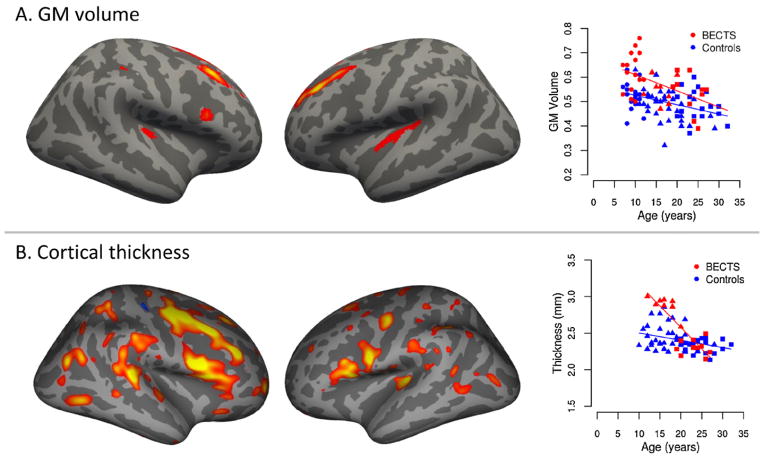
Key findings: The voxel-based morphometry analysis indicated increased bilateral grey matter volume in the superior frontal gyrus, insula and right inferior frontal gyrus regions in BECTS. The magnitude of the increase lessened with age of the cases. Cortical thickness analysis revealed thicker cortex in BECTS along middle and inferior frontal gyri bilaterally, left insula and bilateral supramarginal gyrus in the 9-year-after-onset group, that normalised with age. The rate of cortical thickness changes with age were greater in BECTS cases than in controls.
Significance: Increased cortical gray matter associated with BECTS was found. The decreasing magnitude of the effect with increasing age parallels the natural history of the disorder. The areas affected are consistent with neurocognitive dysfunction in BECTS.
Copyright © 2013 Elsevier B.V. All rights reserved.
Figures
References
-
- Ashburner J, Friston KJ. Voxel-based morphometry—the methods. Neuroimage. 2000;11 (6 Pt 1):805–821. - PubMed
-
- Ashburner J, Friston KJ. Unified segmentation. Neuroimage. 2005;26 (3):839–851. - PubMed
-
- Berg AT, Levy SR, et al. Classification of childhood epilepsy syndromes in newly diagnosed epilepsy: interrater agreement and reasons for disagreement. Epilepsia. 1999;40 (4):439–444. - PubMed
-
- Bouma PA, Bovenkerk AC, et al. The course of benign partial epilepsy of childhood with centrotemporal spikes: a meta-analysis. Neurology. 1997;48 (2):430–437. - PubMed
Publication types
MeSH terms
Grants and funding
LinkOut - more resources
Full Text Sources
Other Literature Sources
Medical
