. 2013 Mar;9(2):99-112.
doi: 10.1016/j.jalz.2012.11.005. Epub 2013 Jan 30.

New ELISAs with high specificity for soluble oligomers of amyloid β-protein detect natural Aβ oligomers in human brain but not CSF

Affiliations

New ELISAs with high specificity for soluble oligomers of amyloid β-protein detect natural Aβ oligomers in human brain but not CSF

Ting Yang et al. Alzheimers Dement. 2013 Mar.

Abstract

Background: Soluble oligomers of amyloid ß-protein (Aß) have been increasingly linked to synaptic dysfunction, tau alteration, and neuritic dystrophy in Alzheimer's disease (AD) and mouse models. There is a great need for assays that quantify Aß oligomers with high specificity and sensitivity.

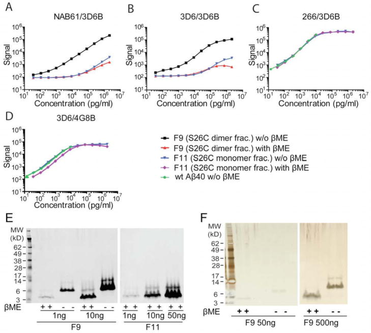
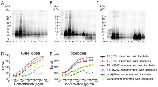
Methods: We designed and validated two oligomer-specific (o-) enzyme-linked immunoassays (ELISAs) using either an Aß aggregate-selective monoclonal for capture and a monoclonal to the free N-terminus for detection, or the latter antibody for both capture and detection.

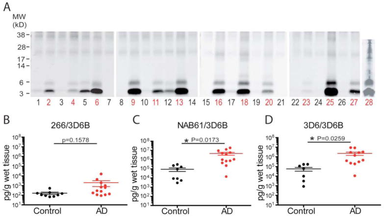
Results: The o-ELISAs specifically quantified pure oligomers of synthetic Aß with sizes from dimers up to much larger assemblies and over a wide dynamic range of concentrations, whereas Aß monomers were undetectable. Natural Aß oligomers of similarly wide size and concentration ranges were measured in extracts of AD and control brains, revealing >1000-fold higher concentrations of Aß oligomers than monomers in the soluble fraction of AD cortex. The assays quantified the age-related rise in oligomers in hAPP transgenic mice. Unexpectedly, none of 90 human cerebrospinal fluid (CSF) samples gave a specific signal in either o-ELISA.

Conclusions: These new o-ELISAs with rigorously confirmed specificity can quantify oligomer burden in human and mouse brains for diagnostic and mechanistic studies and for AD biomarker development. However, our data raise the likelihood that the hydrophobicity of Aß oligomers makes them very low in number or absent in aqueous CSF.


Figures

Figure 1
Figure 1. 3D6/3D6B and NAB61/3D6B ELISAs specifically recognize Aβ dimers but not monomers
Both NAB61/3D6B (A) and 3D6/3D6B (B) show wide dynamic ranges for quantifying SEC-purified Aβ S26C dimer (F9; black squares), but do not detect F9 reduced to monomers by βME (red triangles) or Aβ S26C monomers (SEC F11; blue inverse triangle). Conventional Aβ ELISAs 266/3D6B (C) and 3D6/4G8B (D) detect F11 (blue inverse triangles), β ME-treated F11 (purple diamond), and the wt synthetic Aβ40 (green circles) equally well, indicating that β ME does not interfere. Data are means ± SEMs (error bars too small to see on this log scale). (E, F) βME effectively dissociates the SDS-stable S26C dimers of F9 into monomers, with small amounts of residual dimers (<10%). At high concentrations, the F11 monomer fraction can be seen to have small amounts of dimers. (E: 3D6 WB; F: silver stain).
Figure 2
Figure 2. The o-ELISAs recognize a large size range of synthetic Aβ oligomers, including dimers
(A–C) WB (6E10+2G3+21F12) of the SEC elution profile of F9 (A), F11 (B) and wt synthetic Aβ40 monomer (C) that had each been incubated at 37°C overnight and then subjected to SEC (WB: 6E10+2G3+21F12). (D, E) Samples before and after 37°C incubation were serially diluted and assayed by NAB61/3D6B (D) or 3D6/3D6B (E); data are means ± SEM.
Figure 3
Figure 3. The o-ELISAs detect multiple oligomeric forms of Aβ, including dimers, in soluble extracts of human brain tissue
(A) NAB61/3D6B and (B) 3D6/3D6B o-ELISAs, and (C) 266/3D6B 1-x ELISA. By each method, ~80% of soluble Aβ species elute in the void volume of the Superdex 75 SEC column (i.e., F3–F5, corresponding to >70 kDa). (D) SEC elution profiles of AD-TBS extracts analyzed by WB (6E10+2G3+21F12); synthetic wt Aβ40 (2 ng) is a WB control.
Figure 4
Figure 4. The o-ELISAs specifically detect Aβ species in AD TBS brain extracts
(A–B) AD-TBS was serially diluted and assayed with the two o-ELISAs (A) and the conventional Aβ1-x ELISA (B). All three ELISAs showed highly linear concentration curves. (C) After two sequential pre-clearing steps, AD-TBS was subjected to IP by antibodies 3D6 or 3F3 or NAB61. As numbered, the various supernatants were assayed by o-ELISAs (bar graph) and the corresponding immunoprecipitates by WB. (WB: 6E10+2G3+21F12).
Figure 5
Figure 5. Aqueously soluble Aβ oligomers are significantly increased in AD vs. control brains
(A) IP/WB of TBS extracts (IP: AW8; WB: 6E10+2G3+21F12). Each lane represents an individual subject (black: control; red: AD). (B–D) Scatterplots for total Aβ (1-x) levels (B) and Aβ oligomer levels by NAB61/3D6B (C) or 3D6/3D6B (D) in human brain TBS extracts. Each dot represents data from 1 subject. Horizontal lines: means ± SEMs. p values from two-tailed Mann-Whitney t-tests.
Figure 6
Figure 6. Lack of Aβ oligomer signals in CSF of representative control and AD subjects by IP/WB and o-ELISAs
IP/WB analysis on (A) 2 ml CSF (IP: Aβ antiserum R1282; WB: 6E10+2G3+21F12), and (B) 5 ml CSF (1st IP: 3D6 (3ug/ml), 2nd IP: Aβ antiserum R1282; WB: 6E10+2G3+21F12). Clinical information is in Table 3. (C–D) S26C dimer fraction (F9) was spiked into a human CSF, serially diluted, and assayed by NAB61/3D6B (C) and 3D6/3D6B (D) o-ELISAs. Readings generated from F9 diluted in CSF (red squares) show a perfect overlap with those of F9 diluted in specimen diluent (black circles). Data are means ± SEMs.

References

    1. Motter R, Vigo-Pelfrey C, Kholodenko D, Barbour R, Johnson-Wood K, Galasko D, et al. Reduction of beta-amyloid peptide 42 in the cerebrospinal fluid of patients with Alzheimer’s disease. Ann Neurol. 1995;38:643–648. - PubMed
    1. Fagan AM, Mintun MA, Mach RH, Lee SY, Dence CS, Shah AR, et al. Inverse relation between in vivo amyloid imaging load and cerebrospinal fluid Abeta42 in humans. Ann Neurol. 2006;59:512–519. - PubMed
    1. Fagan AM, Head D, Shah AR, Marcus D, Mintun M, Morris JC, et al. Decreased cerebrospinal fluid Abeta(42) correlates with brain atrophy in cognitively normal elderly. Ann Neurol. 2009;65:176–183. - PMC - PubMed
    1. Shaw LM, Vanderstichele H, Knapik-Czajka M, Clark CM, Aisen PS, Petersen RC, et al. Cerebrospinal fluid biomarker signature in Alzheimer’s disease neuroimaging initiative subjects. Ann Neurol. 2009;65:403–413. - PMC - PubMed
    1. Fagan AM, Roe CM, Xiong C, Mintun MA, Morris JC, Holtzman DM. Cerebrospinal fluid tau/beta-amyloid(42) ratio as a prediction of cognitive decline in nondemented older adults. Arch Neurol. 2007;64:343–349. - PubMed

Publication types

MeSH terms