Review
. 2013 Apr;175(1-2):51-60.
doi: 10.1016/j.autneu.2012.12.009. Epub 2013 Jan 30.

Bidirectional neuro-glial signaling modalities in the hypothalamus: role in neurohumoral regulation

Affiliations
Review

Bidirectional neuro-glial signaling modalities in the hypothalamus: role in neurohumoral regulation

J E Stern et al. Auton Neurosci. 2013 Apr.

Abstract

Maintenance of bodily homeostasis requires concerted interactions between the neuroendocrine and the autonomic nervous systems, which generate adaptive neurohumoral outflows in response to a variety of sensory inputs. Moreover, an exacerbated neurohumoral activation is recognized to be a critical component in numerous disease conditions, including hypertension, heart failure, stress, and the metabolic syndrome. Thus, the study of neurohumoral regulation in the brain is of critical physiological and pathological relevance. Most of the work in the field over the last decades has been centered on elucidating neuronal mechanisms and pathways involved in neurohumoral control. More recently however, it has become increasingly clear that non-neuronal cell types, particularly astrocytes and microglial cells, actively participate in information processing in areas of the brain involved in neuroendocrine and autonomic control. Thus, in this work, we review recent advances in our understanding of neuro-glial interactions within the hypothalamic supraoptic and paraventricular nuclei, and their impact on neurohumoral integration in these nuclei. Major topics reviewed include anatomical and functional properties of the neuro-glial microenvironment, neuron-to-astrocyte signaling, gliotransmitters, and astrocyte regulation of signaling molecules in the extracellular space. We aimed in this review to highlight the importance of neuro-glial bidirectional interactions in information processing within major hypothalamic networks involved in neurohumoral integration.


Figures

Figure 1
Figure 1. Endothelin B (ETB) receptor activation in the SON increases intracellular Ca2+ levels in astrocytes, and evokes a delayed change in SON firing activity
A1, Representative pseudocolor images of transient Ca2+ changes in Fluo-4 loaded astrocytes (arrows) induced by the ETB receptor agonist sarafotoxin (S6c). A2, traces of Δ Ca2+ (ΔF/F0) corresponding to the astrocytes shown in A1. A3, Summary data of ΔF/F0 in response to S6c in astrocytes loaded with Fluo4-AM or Rhod2-AM. B, Bath application of Sarafotoxin 6c (S6c, 100 nM) induced either excitatory (B1) or inhibitory (B2) responses in SON neurons. Pre-incubation of hypothalamic slices in the presence of the gliotoxin L-αAA (250 μM) prevented S6c effects on SON firing activity (B3). ***P< 0.001. Scale bar= 10 μm. Modified from (Filosa et al., 2012).
Figure 2
Figure 2. Glial eNOS modulates presympathetic neuronal activity and renal sympathetic nerve outflow in the PVN
A and B: Immunostaining of eNOS (green) and the glial marker GFAP (red) within the PVN. B: Higher magnification image of the area outlined in A. C and D: eNOS (green) and glial S100b (red) immunoreactivities within the PVN. D: Higher magnification image of the area outlined by the square in C. Note the dense eNOS staining in the PVN, and the colocalization (yellow color: green + red) with both glial markers. EG: Photomicrographs showing staining for the nitric oxide sensitive dye DAF-2 under (E) basal condition (ACSF), or in the presence of (F) eNOS inhibitor L-NIO (10μmol/L), and (G) eNOS inhibitor Cavtratin (10μmol/L). H: Summary data showing mean DAF-2 intensity in each experimental condition. I: in vitro electrophysiological recordings sowing firing activity of a PVN-RVLM projecting neuron before (top), during (middle) and after (bottom) bath application of L-NIO (10μmol/L). J: Summary data for mean firing frequency in PVN-RVLM neurons. K: Dose-dependent changes in renal sympathetic nerve activity (RSNA) in response to microinjections of L-NIO (50, 100, and 200pmol) into the PVN. Scale bars: 50μm. 3V: third ventricle; *P<0.05 vs *ACSF (H and J). Modified from (Biancardi et al., 2011).
Figure 3
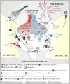
Figure 3. Schematic diagram depicting the various signals and targets at the SON/PVN tripartite synapse

Similar articles

Cited by

References

    1. Agulhon C, Petravicz J, McMullen AB, Sweger EJ, Minton SK, Taves SR, Casper KB, Fiacco TA, McCarthy KD. What is the role of astrocyte calcium in neurophysiology? Neuron. 2008;59:932–946. - PMC - PubMed
    1. Akine A, Montanaro M, Allen AM. Hypothalamic paraventricular nucleus inhibition decreases renal sympathetic nerve activity in hypertensive and normotensive rats. Auton Neurosci. 2003;108:17–21. - PubMed
    1. Allen AM. Inhibition of the hypothalamic paraventricular nucleus in spontaneously hypertensive rats dramatically reduces sympathetic vasomotor tone. Hypertension. 2002;39:275–280. - PubMed
    1. Araque A, Navarrete M. Glial cells in neuronal network function. Philos Trans R Soc Lond B Biol Sci. 2010;365:2375–2381. - PMC - PubMed
    1. Araque A, Parpura V, Sanzgiri RP, Haydon PG. Tripartite synapses: glia, the unacknowledged partner. Trends Neurosci. 1999;22:208–215. - PubMed

Publication types

LinkOut - more resources