

Somatic uniparental isodisomy explains multifocality of glomuvenous malformations
- PMID: 23375657
- PMCID: PMC3567282
- DOI: 10.1016/j.ajhg.2012.12.017
Somatic uniparental isodisomy explains multifocality of glomuvenous malformations
Abstract
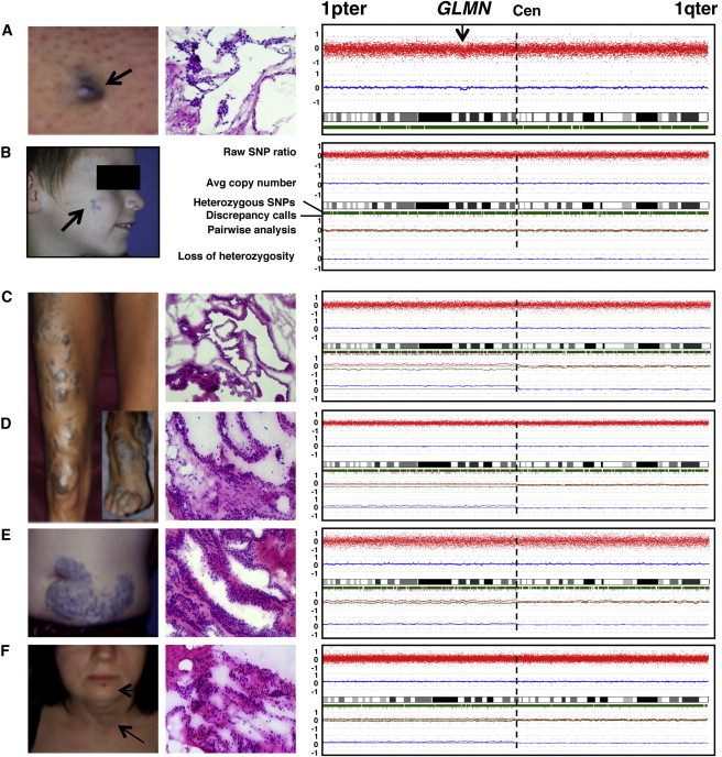
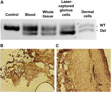
Inherited vascular malformations are commonly autosomal dominantly inherited with high, but incomplete, penetrance; they often present as multiple lesions. We hypothesized that Knudson's two-hit model could explain this multifocality and partial penetrance. We performed a systematic analysis of inherited glomuvenous malformations (GVMs) by using multiple approaches, including a sensitive allele-specific pairwise SNP-chip method. Overall, we identified 16 somatic mutations, most of which were not intragenic but were cases of acquired uniparental isodisomy (aUPID) involving chromosome 1p. The breakpoint of each aUPID is located in an A- and T-rich, high-DNA-flexibility region (1p13.1-1p12). This region corresponds to a possible new fragile site. Occurrences of these mutations render the inherited glomulin variant in 1p22.1 homozygous in the affected tissues without loss of genetic material. This finding demonstrates that a double hit is needed to trigger formation of a GVM. It also suggests that somatic UPID, only detectable by sensitive pairwise analysis in heterogeneous tissues, might be a common phenomenon in human cells. Thus, aUPID might play a role in the pathogenesis of various nonmalignant disorders and might explain local impaired function and/or clinical variability. Furthermore, these data suggest that pairwise analysis of blood and tissue, even on heterogeneous tissue, can be used for localizing double-hit mutations in disease-causing genes.
Copyright © 2013 The American Society of Human Genetics. Published by Elsevier Inc. All rights reserved.
Figures
References
-
- Boon L.M., Mulliken J.B., Enjolras O., Vikkula M. Glomuvenous malformation (glomangioma) and venous malformation: Distinct clinicopathologic and genetic entities. Arch. Dermatol. 2004;140:971–976. - PubMed
-
- Goodman T.F., Abele D.C. Multiple glomus tumors. A clinical and electron microscopic study. Arch. Dermatol. 1971;103:11–23. - PubMed
-
- Irrthum A., Brouillard P., Enjolras O., Gibbs N.F., Eichenfield L.F., Olsen B.R., Mulliken J.B., Boon L.M., Vikkula M. Linkage disequilibrium narrows locus for venous malformation with glomus cells (VMGLOM) to a single 1.48 Mbp YAC. Eur. J. Hum. Genet. 2001;9:34–38. - PubMed
Publication types
MeSH terms
Substances
Supplementary concepts
Grants and funding
LinkOut - more resources
Full Text Sources
Other Literature Sources
Molecular Biology Databases
