. 2013 Aug 1;74(3):212-9.
doi: 10.1016/j.biopsych.2012.12.013. Epub 2013 Jan 31.

Default mode network in childhood autism: posteromedial cortex heterogeneity and relationship with social deficits

Affiliations

Default mode network in childhood autism: posteromedial cortex heterogeneity and relationship with social deficits

Charles J Lynch et al. Biol Psychiatry. .

Abstract

Background: The default mode network (DMN), a brain system anchored in the posteromedial cortex, has been identified as underconnected in adults with autism spectrum disorder (ASD). However, to date there have been no attempts to characterize this network and its involvement in mediating social deficits in children with ASD. Furthermore, the functionally heterogeneous profile of the posteromedial cortex raises questions regarding how altered connectivity manifests in specific functional modules within this brain region in children with ASD.

Methods: Resting-state functional magnetic resonance imaging and an anatomically informed approach were used to investigate the functional connectivity of the DMN in 20 children with ASD and 19 age-, gender-, and IQ-matched typically developing (TD) children. Multivariate regression analyses were used to test whether altered patterns of connectivity are predictive of social impairment severity.

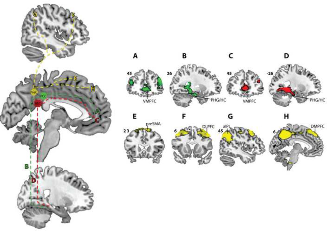
Results: Compared with TD children, children with ASD demonstrated hyperconnectivity of the posterior cingulate and retrosplenial cortices with predominately medial and anterolateral temporal cortex. In contrast, the precuneus in ASD children demonstrated hypoconnectivity with visual cortex, basal ganglia, and locally within the posteromedial cortex. Aberrant posterior cingulate cortex hyperconnectivity was linked with severity of social impairments in ASD, whereas precuneus hypoconnectivity was unrelated to social deficits. Consistent with previous work in healthy adults, a functionally heterogeneous profile of connectivity within the posteromedial cortex in both TD and ASD children was observed.

Conclusions: This work links hyperconnectivity of DMN-related circuits to the core social deficits in young children with ASD and highlights fundamental aspects of posteromedial cortex heterogeneity.

Keywords: Autism spectrum disorders; default mode network; functional connectivity; posterior cingulate cortex; posteromedial cortex; resting-state fMRI.


Conflict of interest statement

Financial Disclosures: All of the authors have reported no biomedical financial interests or potential conflicts of interest.

Figures

Figure 1
Figure 1
Summary of posteromedial cortex anatomy and connectivity based on the work of Margulies and colleagues (25). The posteromedial cortex (PMC) (A) encompasses the posterior cingulate cortex (PCC), retrosplenial cortex (RSC), and precuneus (PreC) (B). Ventral aspects of the PMC, including the PCC (C) and RSC (D), have strong connections with medial temporal lobe (MTL) and ventromedial prefrontal cortex (VMPFC). The PreC (E) has stronger connections with dorsolateral prefrontal cortex (DLPFC), supplementary motor (SMA), and occipital regions. cc = corpus callosum, cs = cingulate sulcus, pof = parieto-occipital sulcus, sps = sub-parietal sulcus.
Figure 2
Figure 2
Posteromedial cortex ventral and dorsal sub-regions demonstrate differential profiles of connectivity. Posterior cingulate and retrosplenial cortices demonstrated stronger connections with VMPFC and medial temporal lobe DMN nodes compared to the precuneus (A, B, C, D). The precuneus was most strongly connected with the pre-supplementary motor area (preSMA) (E), dorsolateral prefrontal cortex (DLPFC) (F), anterior inferior parietal lobule (aIPL) (G), and dorsomedial aspects of the medial prefrontal cortex (DMPFC) (H).
Figure 3
Figure 3
Children with ASD demonstrated both hyper-connectivity (ASD > TD) and hypo-connectivity (TD > ASD) of posteromedial cortex sub-regions. The posterior cingulate cortex (PCC) and retrosplenial cortex (RSC) revealed ASD hyper-connectivity while the precuneus (PreC) demonstrated ASD hypo-connectivity. *p < 0.01, **p < 0.005, ***p < 0.001. aLTC = anterolateral temporal cortex, DMPFC = dorsomedial prefrontal cortex, ERc = entorhinal cortex, LG = lingual gyrus, PHG = parahippocampal gyrus, pInsula = posterior insular cortex, PRc = perirhinal cortex, pSTS = posterior superior temporal sulcus, TempP = temporal pole.
Figure 4
Figure 4
Posterior cingulate cortex hyper-connectivity predicts social deficits in ASD. Connections between the posterior cingulate cortex (PCC) seed region of interest (ROI) and associated ASD hyper-connected target regions were found to be predictive of social impairments as measured by the ADOS social subscale (A). No such significant relationships were demonstrated by the retrosplenial cortex (RSC) and precuneus (PreC)(B). L= left, R= right, n.s. = not significant, * represents significance. PHG = parahippocampal gyrus, TempP = temporal pole, LG = lingual gyrus.

References

    1. Baio J. Prevalence of autism spectrum disorders - autism and developmental disabilities monitoring network, 14 sites, United States, 2008. MMWR Surveill Summ. 2012;61:1–19. - PubMed
    1. Lombardo MV, Barnes JL, Wheelwright SJ, Baron-Cohen S. Self-referential cognition and empathy in autism. PLoS ONE. 2007;2:e883. - PMC - PubMed
    1. Uddin LQ. The self in autism: an emerging view from neuroimaging. Neurocase. 2011;17:201–208. - PMC - PubMed
    1. Greicius MD, Krasnow B, Reiss AL, Menon V. Functional connectivity in the resting brain: a network analysis of the default mode hypothesis. Proc Natl Acad Sci U S A. 2003;100:253–258. - PMC - PubMed
    1. Raichle ME, MacLeod AM, Snyder AZ, Powers WJ, Gusnard DA, Shulman GL. A default mode of brain function. Proc Natl Acad Sci U S A. 2001;98:676–682. - PMC - PubMed

Publication types

LinkOut - more resources