Review
. 2013 Nov:74:4-17.
doi: 10.1016/j.neuropharm.2013.01.016. Epub 2013 Jan 31.

Influence of GluN2 subunit identity on NMDA receptor function

Affiliations
Review

Influence of GluN2 subunit identity on NMDA receptor function

D J A Wyllie et al. Neuropharmacology. 2013 Nov.

Abstract

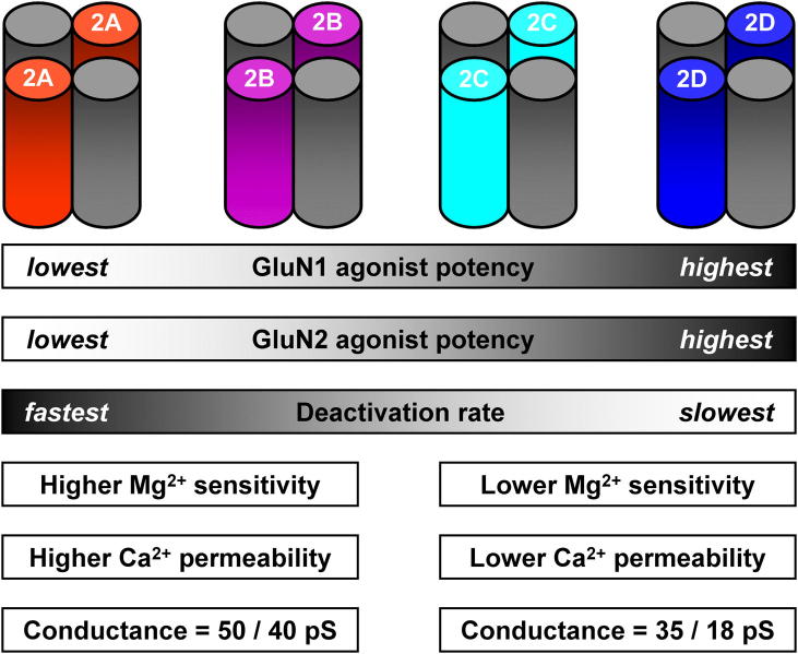
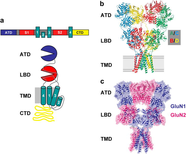
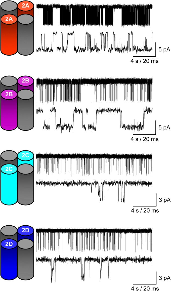
N-methyl-d-aspartate receptors (NMDARs) are ligand-gated ion channels ('ionotropic' receptors) activated by the major excitatory neurotransmitter, l-glutamate. While the term 'the NMDAR' is often used it obscures the fact that this class of receptor contains within it members whose properties are as different as they are similar. This heterogeneity was evident from early electrophysiological, pharmacological and biochemical assessments of the functional properties of NMDARs and while the molecular basis of this heterogeneity has taken many years to elucidate, it indicated from the outset that the diversity of NMDAR phenotypes could allow this receptor family to subserve a variety of functions in the mammalian central nervous system. In this review we highlight some recent studies that have identified structural elements within GluN2 subunits that contribute to the heterogeneous biophysical properties of NMDARs, consider why some recently described novel pharmacological tools may permit better identification of native NMDAR subtypes, examine the evidence that NMDAR subtypes differentially contribute to the induction of long-term potentiation and long-term depression and discuss how through the use of chimeric proteins additional insights have been obtained that account for NMDAR subtype-dependency of physiological and pathophysiological signalling. This article is part of the Special Issue entitled 'Glutamate Receptor-Dependent Synaptic Plasticity'.

Keywords: Biophysics; Excitoxicity; Glutamate receptor; NMDA receptor; Pharmacology; Plasticity; Structure.


Figures

Fig. 1
Fig. 1
The identity of the GluN2 subunit controls pharmacological and biophysical properties of NMDARs. Schematic representation of each of the four di-heteromeric NMDARs with the GluN1 subunits indicated in grey. Agonist and co-agonist potency is lowest at GluN1/GluN2A NMDARs and highest at GluN1/GluN2D NMDARs. Deactivation rates are fastest for GluN2A-containing NMDARs and slowest for NMDARs containing GluN2D subunits. GluN2B- and GluN2C-containing NMDARs show approximately similar deactivation rates. NMDARs fall into two categories (GluN2A/B-like and GluN2C/D-like) when considering voltage-dependent block by Mg2+, permeability to Ca2+ and unitary conductance.
Fig. 2
Fig. 2
Structure of ionotropic glutamate receptors. (a) Upper panel, linear representation of iGluR subunit highlighting the four functional domains; lower panel, schematic of the general structure of an iGluR subunit indicating the extracellularly located amino terminal domain (ATD) and ligand-binding domain (LBD) the transmembrane domain (TMD) comprised of three membrane-spanning helices (M1, M3 and M4) together with the re-entrant P-loop region of M2 and the large intracellularly located C-terminal domain (CTD). (b) Ribbon structure representation of the rat GluA2 homomeric AMPAR with each of the four subunits coloured coded and indicating the two conformationally distinct pairs of subunits, A/C and B/D. (c) model of the overall architecture of the NMDAR based on the GluA2 crystal structures of the ATD and TMD and the GluN1-GluN2A LBD heterodimer crystal structure. Panels (b) and (c) adapted from Sobolevsky et al. (2009). Copyright © 2009 Nature Publishing Group. Used with permission.
Fig. 3
Fig. 3
The Ser/Leu site in M3 controls NMDAR single-channel conductance (γ) and voltage-dependent block by Mg2+. (a) The characteristic high (∼50 pS) conductance of GluN1/GluN2A NMDARs is changed to a GluN1/GluN2D-like conduction by the point mutation GluN2A(S632L). (b) The reciprocal mutation in GluN2D that replaces the leucine residue at position 657 by serine gives rise to single-channel currents with a GluN1/GluN2A-like conductance. (c) The IC50 values for voltage-dependent Mg2+-block for GluN1/GluN2A(S632L) are comparable to those for GluN1/GluN2D NMDARs while IC50 values for GluN1/GluN2D(L657S) resemble those for GluN1/GluN2A NMDARs. Panels (a)-(c) from Siegler Retchless et al. (2012). Copyright © 2012 Nature Publishing Group. Used with permission.
Fig. 4
Fig. 4
The single-channel characteristics of NMDARs are determined by the GluN2 subunit assembled within the receptor. Steady-state single-channel recordings from excised outside-out membrane patches isolated from HEK293 cells expressing either GluN1/GluN2A, GluN1/GluN2B, GluN1/GluN2C, or GluN1/GluN2D NMDARs. For each receptor combination the upper panel illustrates 20 s of activity while the lower panel shows a selected higher resolution 100 ms period of activity. For all recordings, channel activity was elicited by glutamate (1 mM) in the presence of glycine (0.05 mM) and performed at pH 8.0 in 0.5 mM Ca2+. Unpublished data, kindly provided by S. M. Dravid, K. Erreger, K. Ogden, K. M. Vance, and S. F. Traynelis.
Fig. 5
Fig. 5
Control of NMDAR deactivation rates. (a) Example of a burst of openings arising from an activation of a wild-type (WT) GluN1/GluN2D NMDAR (ai. upper panel, grey); lower three traces (black) show examples of single activations of GluN1/GluN2D(T692A) NMDARs. For each the line denotes the period between the first opening and last closing of each burst. Comparison of burst length distributions (aii) for GluN1/GluN2D(T692) and WT GluN1/GluN2D NMDARs (shown as a dashed grey line). Note the numbering used to indicate the position of the threonine residue in GluN2D is for the mature protein (i.e. it excludes the signal peptide). (b) Representative whole-cell currents recorded from an HEK293 cell expressing GluN1/GluN2D NMDARs (bi) or GluN1/GluN2D NMDARs in which the ATD was removed (bii). Note the increase in the deactivation rate when the GluN2D ATD is absent. (c) Exon-5 lacking GluN1-1a subunits (ci) when co-expressed with GluN2D form NMDARs with typical slow deactivation rates, whereas GluN1-1b subunits which containing exon 5 (cii) accelerate deactivation of GluN2D-containing NMDARs. Panel (a) from Chen et al. (2004); (b) from Yuan et al. (2009) and (c) from Vance et al. (2012). Used with permission.
Fig. 6
Fig. 6
Antagonism of NMDAR-mediated currents by TCN 201. (a) Left, example steady-state whole-cell NMDAR-mediated currents recorded from cortical pyramidal cells from (ai), days in vitro (DIV) 9–10 neurones, (aii), DIV 9–10 neurones transfected with GluN2A NMDAR subunits, and (aiii), DIV 15–18 neurones. To the right, traces illustrate the sensitivity of each of these NMDAR-mediated currents to the GluN2B-selective antagonist, ifenprodil and the subsequent sensitivity of the ifenprodil-insensitive component of this current to TCN 201. (b) Plot illustrating the extent of ifenprodil and TCN 201 antagonism of NMDA-evoked currents. Despite a wide range in the amount of block produced by either ifenprodil or TCN 201 the data show a strong (negative) correlation. (c) Equivalent plot to that illustrated in (b) but for antagonism by ifenprodil and TCN 213. Panels (a) and (b) from Edman et al. (2012) and (c) from McKay et al. (2012). Used with permission.
Fig. 7
Fig. 7
The identity of the GluN2 CTD determines the response to excitotoxic insult. (a) Cartoon depiction illustrating the C-terminal replacement (CTR) of the GluN2B CTD with that from GluN2A and denoted as GluN2B2A(CTR). (b) Quantification of hippocampal lesion volumes 24 h after stereotaxic injection of phosphate buffered saline (PBS) or 15 nmol of NMDA into the hippocampi of either GluN2B+/+ or GluN2B2A(CTR)/2A(CTR) mice. Note the significant reduction in lesion volumes when the GluN2B CTD is replaced by that of GluN2A. (c) Upper panels, representative pictures of haematoxylin and eosin-stained hippocampal sections showing the extent of NMDA-induced damage (dashed lines); lower panels, higher magnification of the boxed areas shown in the upper panels. Calibration bars in upper and lower panels are 250 μm and 50 μm, respectively. Panels (b) and (c) from Martel et al. (2012). Copyright © 2012 Cell Press. Used with permission.

Similar articles

Cited by

References

    1. Aarts M., Liu Y., Liu L., Besshoh S., Arundine M., Gurd J.W., Wang Y.T., Salter M.W., Tymianski M. Treatment of ischemic brain damage by perturbing NMDA receptor- PSD-95 protein interactions. Science. 2002;298:846–850. - PubMed
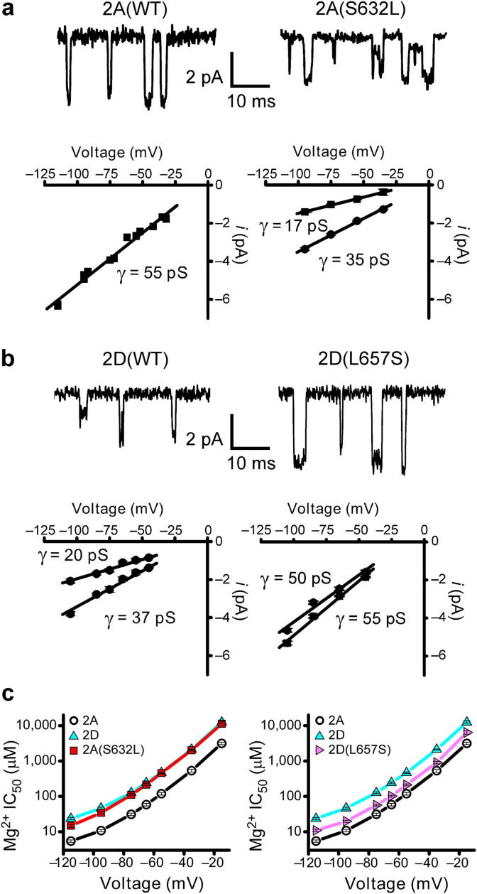
    1. Acker T.M., Yuan H., Hansen K.B., Vance K.M., Ogden K.K., Jensen H.S., Burger P.B., Mullasseril P., Snyder J.P., Liotta D.C., Traynelis S.F. Mechanism for noncompetitive inhibition by novel GluN2C/D N-methyl-d-aspartate receptor subunit-selective modulators. Mol. Pharmacol. 2011;80:782–795. - PMC - PubMed
    1. Al-Mubarak B., Soriano F.X., Hardingham G.E. Synaptic NMDAR activity suppresses FOXO1 expression via a cis-acting FOXO binding site: FOXO1 is a FOXO target gene. Channels (Austin) 2009;3:233–238. - PMC - PubMed
    1. Amico-Ruvio S.A., Popescu G.K. Stationary gating of GluN1/GluN2B receptors in intact membrane patches. Biophys. J. 2010;98:1160–1169. - PMC - PubMed
    1. Anson L.C., Chen P.E., Wyllie D.J., Colquhoun D., Schoepfer R. Identification of amino acid residues of the NR2A subunit that control glutamate potency in recombinant NR1/NR2A NMDA receptors. J. Neurosci. 1998;18:581–589. - PMC - PubMed

Publication types

MeSH terms

Substances