. 2013 Apr 15;532(2):61-72.
doi: 10.1016/j.abb.2013.01.006. Epub 2013 Jan 29.

Oncogenic Ras suppresses Cdk1 in a complex manner during the incubation of activated Xenopus egg extracts

Affiliations

Oncogenic Ras suppresses Cdk1 in a complex manner during the incubation of activated Xenopus egg extracts

Tun-Lan Huang et al. Arch Biochem Biophys. .

Abstract

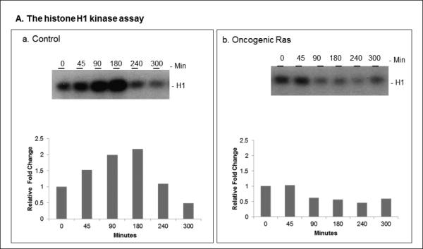
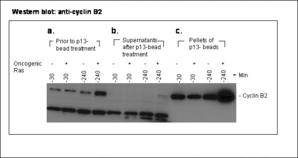
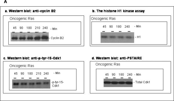
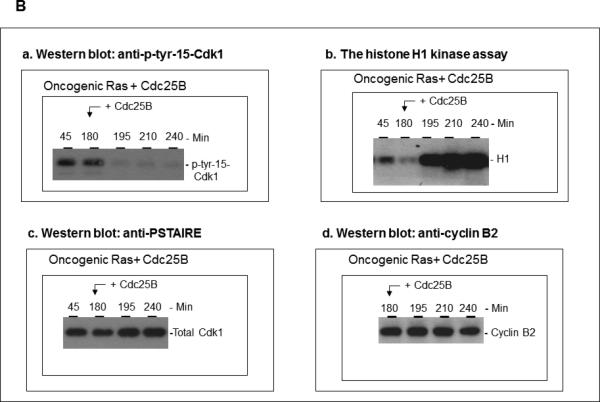
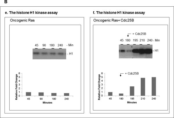
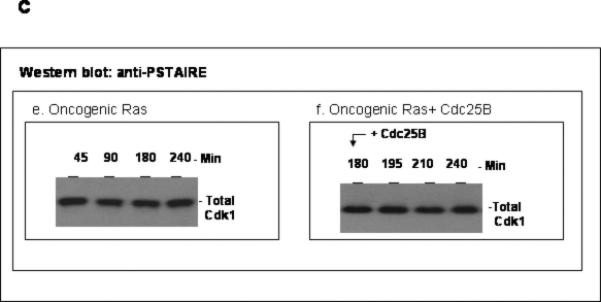
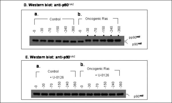
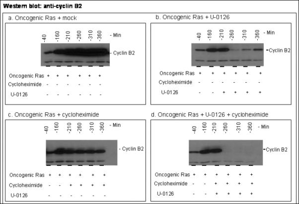
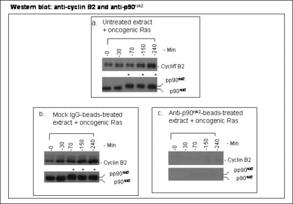
The activity of Cdk1 is the driving force for entry into M-phase during the cell cycle. Activation of Cdk1 requires synthesis and accumulation of cyclin B, binding of cyclin B to Cdk1, and removal of the inhibitory tyr-15-Cdk1 phosphorylation. It was previously shown that oncogenic Ras suppresses Cdk1 activation during the incubation of activated Xenopus egg extracts. However, how oncogenic Ras suppresses Cdk1 remained unclear. Using the histone H1 kinase assay to follow Cdk1 activity and Western blot analysis to assess levels of both cyclin B2 and phosphorylated-tyr-15-Cdk1, how oncogenic Ras suppresses Cdk1 is studied. The results indicate that oncogenic Ras suppresses Cdk1 via induction of persistent phosphorylation of tyr-15-Cdk1. Interestingly, the results reveal that, compared with cyclin B2 in control activated egg extracts, which increased, peaked and then declined during the incubation, oncogenic Ras induced continuous accumulation of cyclin B2. The results also indicate that oncogenic Ras induces continuous accumulation of cyclin B2 primarily through stabilization of cyclin B2, which is mediated by constitutive activation of the Raf-Mek-Erk-p90(rsk) pathway. Taken together, these results indicate that oncogenic Ras suppresses Cdk1 in a complex manner: It induces continuous accumulation of cyclin B2, but also causes persistent inhibitory phosphorylation of tyr-15-Cdk1.


Figures

Figure 1
Figure 1. Oncogenic Ras induces continuous accumulation of cyclin B2 but suppresses the activation of the histone H1 kinase (Cdk1) during the incubation of activated egg extracts
To two 400 μl aliquots of an activated egg extract, either a buffer solution containing 1 mM DTT and 10 mM Hepes, pH 7.8, or a purified, bacterially expressed human oncogenic RasH in the buffer solution (final ~5 μg/ml extract) was added respectively (designated as the control extract or the oncogenic Ras extract, respectively), and the aliquots were then incubated at room temperature, as described in the Materials and Methods. Samples were collected during the incubation and subjected to both the histone H1 kinase assay (1A) and Western blot analysis using anti-cyclin B2 antibody (X121) (1B) and anti-PSTAIRE antibody (1C), as described in the Materials and Methods. Autoradiograms: Panels a of 1A, 1B and 1C, the control extract; panels b of 1A, 1B and 1C, the oncogenic Ras extract. Conditions are indicated on each panel. Note that in 1A, the relative density of H1 bands to the background in each autoradiogram was also quantitated as described in the Materials and Methods. The values were normalized to the value of the initial time-point of the autoradiogram, and expressed as relative fold change in bar graphs.
Figure 2
Figure 2. Cyclin B2 binds to p13suc1-agarose beads
Two aliquots of an activated egg extract were incubated at room temperature in either the absence or the presence of oncogenic Ras (designated as the control extract or the oncogenic Ras extract, respectively) as described in Figure 1. Aliquots were collected at 30 minutes and 240 minutes during the incubation, diluted 20-fold with a buffer, and then treated with p13suc1-beads as described in the Materials and Methods. After the treatment, the p13suc1-beads were precipitated by centrifugation. Equal volumes of samples from the diluted extracts prior to p13suc1-bead treatment (panel a) and from the supernatants after p13suc1-bead treatment and centrifugation (panel b), and equal volumes of the eluates from pellets of the p13suc1-beads (panel c) were subjected to SDS-PAGE and Western blot analysis using anti-cyclin B2 antibody (X121), as described in the Materials and Methods. Autoradiogram: Conditions are indicated on each panel. The band of cyclin B2 is indicated. Almost all of the cyclin B2 in both the diluted control extract and the diluted oncogenic Ras extract prior to the p13suc1-bead treatment is removed by p13suc1-beads (compare panel b with panel a). Note that the bands below cyclin B2 in panels a and b are non-specific.
Figure 3
Figure 3. Oncogenic Ras suppresses the histone H1 kinase (Cdk1) via persistent phosphorylation of tyr-15-Cdk1
(3A) The suppression of the histone H1 kinase is correlated with persistent phosphorylation of tyr-15-Cdk1. A 200 μl aliquot of an activated egg extract was incubated at room temperature in the presence of oncogenic Ras (final ~ 5 μg/ml extract) (designated as the oncogenic Ras extract). Samples were collected and subjected to the histone H1 kinase assay and Western blot analysis using anti-cyclin B2 (X121), anti-PSTAIRE and anti-p-tyr-15-Cdk1 antibodies. Autoradiograms: Conditions are indicated on each panel. (3B, 3C) The addition of a constitutively active Cdc25B dephosphorylates p-tyr-15-Cdk1 and abruptly activates the histone H1 kinase. (3B) A portion of the oncogenic Ras extract described in 3A was collected at 180 minutes during the room temperature incubation. Then a constitutively active Cdc25B (final ~10 μg/ml extract) was added to this portion (designated as the oncogenic Ras + Cdc25B extract), which was then further incubated at room temperature. Samples were collected and subjected to both the histone H1 kinase assay and Western blot analysis as described in 3A. Autoradigrams: Conditions are indicated on each panel. Note that Figure 3B (panels e and f) are the same results as Figure 3A (panel b) and Figure 3B (panel b), respectively. In Figure 3B (panels e and f), the relative density of H1 bands to the background in each autoradiogram was quantitated. The values were normalized to the value of the initial time-point in the autoradiogram, and expressed as relative fold change in bar graphs. Also note that the addition of the Cdc25B to the oncogenic Ras extract dephosphorylates p-tyr-15-Cdk1 (compare panel a in 3B with panel c in 3A) and abruptly activates the histone H1 kinase (compare panel b in 3B with panel b in 3A). (3C) An aliquot from another activated egg extract was similarly incubated in the presence of oncogenic Ras as described in 3A. A portion of the oncogenic Ras extract was collected at 180 minutes during the incubation and constitutively active Cdc25B was added to this portion (designated as the oncogenic Ras + Cdc25 B extract) as described in 3B. The oncogenic Ras + Cdc25B extract was further incubated at room temperature. Samples collected from the oncogenic Ras + Cdc25B extract were similarly analyzed as described in 3B. Autoradiograms: Conditions are indicated on each panel. Note that like the results in Figure 3B, the addition of the Cdc25B to the oncogenic Ras extract also dephosphorylates p-tyr-15-Cdk1 (3C; compare panel b with panel a) and abruptly activates the histone H1 kinase (3C; compare panel d with panel c). The total Cdk1 is unchanged (3C, panels e and f). It should also be noted that the experiments for 3C are originally a part of the experiments with Figure 1. The autoradiograms of Figure 3C (panel c and panel e), which show the histone H1 kinase activity and total Cdk1 level during the incubation of the oncogenic Ras extract, are from Figure 1A (panel b) and Figure 1C (panel b), respectively.
Figure 4
Figure 4. The effects of cycloheximide and U-0126 on cyclin B2 levels in activated egg extracts incubated either in the absence or presence of oncogenic Ras
(4A) Oncogenic Ras induces continuous accumulation of cyclin B2. Two 400 μl aliquots of an activated egg extract were incubated at room temperature either in the absence (the control extract) or the presence of oncogenic Ras (the oncogenic Ras extract) as described in Figure 1. Samples were collected and subjected to Western blot analysis using anti-cyclin B2 (X-121). Autoradiograms: Panel a, the control extract; panel b, the oncogenic Ras extract. Conditions are indicated on each panel. The band of cyclin B2 is indicated. Note that the band below cyclin B2 is a non-specific band, and its level does not significantly change during the incubation. (4B) The effect of cycloheximide on cyclin B2 levels: Oncogenic Ras stabilizes cyclin B2 in the presence of cycloheximide. Cycloheximide (final 150 μM) was added to a 100 μl aliquot that was collected from either the control extract or the oncogenic Ras extract described in 4A at 30 minutes during the room temperature incubation. Both the cycloheximide-containing aliquot of the control extract (panel a) and the cycloheximide-containing aliquot of the oncogenic Ras extract (panel b) were further incubated at room temperature. Samples were collected and subjected to Western blot analysis using anti-cyclin B2 (X121). Autoradiograms: Conditions are indicated on each panel. The band of cyclin B2 is indicated. Note that cyclin B2 synthesized prior to the addition of cycloheximide is stable in the oncogenic Ras extract but not in the control extract (compare panel b with panel a). The band below cyclin B2 is a non-specific band, and its level does not significantly change during the incubation. (4C) The effect of U-0126 on cyclin B2 levels: U-0126 dramatically reduces cyclin B2 accumulation in the oncogenic Ras extract. U-0126 (final 50 μM) was added to another 100 μl aliquot that was also collected from either the control extract or the oncogenic Ras extract described in 4A at 30 minutes during the room temperature incubation. Both the U-0126-containing aliquot of the control extract (panel a) and the U-0126-containing aliquot of the oncogenic Ras extract (panel b) were further incubated at room temperature. Samples were collected and subjected to Western blot analysis using anti-cyclin B2 (X121). Autoradiograms: Conditions are indicated on each panel. The band of cyclin B2 is indicated. Note that the addition of U-0126 to the oncogenic Ras extract nearly completely eliminates the oncogenic Ras-induced accumulation of cyclin B2 (compare panel b of 4C with panel b of 4A). The band below cyclin B2 is a non-specific band, and its level does not significantly changed during the incubation. (4D and 4E) Oncogenic Ras induces mobility up-shift of p90rsk2 and U-0126 blocks the oncogenic Ras-induced mobility up-shift. Samples from 4A and 4C were also subjected to Western blot analysis using an anti-p90rsk2 antibody. Autoradiograms: The conditions for 4D and 4E were as Figure 4A and 4C, respectively. Bands of p90rsk2 and pp90rsk2 (up-shifted p90rsk2) are indicated. Lanes with pp90rsk2 are indicated by asterisks.
Figure 5
Figure 5. U-0126 completely blocks the oncogenic Ras-induced stabilization of cyclin B2
Four 100 μl aliquots of an activated egg extract were incubated in the presence of oncogenic Ras (final ~5 μg/ml extract) (designated as the oncogenic Ras extract) as described in Figure 1. Samples were collected at 40 min and 160 min. After sampling, mock (a buffer containing 1 mM DTT and 10 mM Hepes, pH 7.8), U-0126 (final 50 μM), cycloheximide (final 150 μM), or both U-0126 (final 50 μM) and cycloheximide (final 150 μM) was respectively added to the remaining portions of the four aliquots of the oncogenic Ras extract (designated as the oncogenic Ras + mock extract, the oncogenic Ras + U-0126 extract, the oncogenic Ras + cycloheximide extract, and the oncogenic Ras + U-0126 + cycloheximide extract, respectively). The aliquots were then further incubated at room temperature. Samples were collected and subjected to Western blot analysis using anti-cyclin B2 antibody (X121). Autoradiograms: Panel a, the oncogenic Ras + mock extract; panel b, the oncogenic Ras + U-0126 extract; panel c, the oncogenic Ras + cycloheximide extract; and panel d, the oncogenic Ras + U-0126 + cycloheximide extract. Conditions are indicated on each panel. The band of cyclin B2 is indicated. Note that the co-addition of U-0126 and cycloheximide to the oncogenic Ras extract completely abolishes the oncogenic Ras-induced stabilization of cyclin B2 (compare panel d with panel c) and eliminates all cyclin B2 accumulation in the oncogenic Ras extract (compare panel d with panel a). The band below cyclin B2 is a non-specific band, and its level does not significantly change during the incubation.
Figure 6
Figure 6. Immunodepletion of p90rsk2 from activated egg extracts prevents oncogenic Ras from inducing cyclin B2 accumulation
Immunodepletion of p90rsk2 from activated egg extracts was performed as described in the Materials and Methods. Untreated, mock IgG-beads-treated and anti-p90rsk2-beads-treated activated egg extracts were incubated at room temperature in the presence of oncogenic Ras. Samples were collected and subjected to Western blot analysis using anti-p90rsk2 and anti-cyclin B2 (X121) antibodies. Autoradiograms: Conditions are indicated on each panel. The bands of cyclin B2, p90rsk2 and pp90rsk2 are indicated; lanes with pp90rsk2 are indicated by asterisks.

Similar articles

Cited by

References

    1. Malumbres M, Barbacid M. RAS oncogenes: the first 30 years. Nat Rev Cancer. 2003;3:459–465. - PubMed
    1. Land H, Parada LF, Weinberg RA. Tumorigenic conversion of primary embryo fibroblasts requires at least two cooperating oncogenes. Nature. 1983;304:596–602. - PubMed
    1. Hirakawa T, Ruley HE. Rescue of cells from ras oncogene-induced growth arrest by a second, complementing, oncogene. Proc Natl Acad Sci U. S. A. 1988;85:1519–1523. - PMC - PubMed
    1. Ridley AJ, Paterson HF, Noble M, Land H. Ras-mediated cell cycle arrest is altered by nuclear oncogenes to induce Schwann cell transformation. EMBO J. 1988;7:1635–1645. - PMC - PubMed
    1. Lloyd AC. Ras versus cyclin-dependent kinase inhibitors. Curr Opin Genet Dev. 1988;8:43–48. - PubMed

Publication types