. 2013 Mar 22;288(12):8468-8478.
doi: 10.1074/jbc.M112.433540. Epub 2013 Feb 2.

A manganese-rich environment supports superoxide dismutase activity in a Lyme disease pathogen, Borrelia burgdorferi

Affiliations

A manganese-rich environment supports superoxide dismutase activity in a Lyme disease pathogen, Borrelia burgdorferi

J Dafhne Aguirre et al. J Biol Chem. .

Abstract

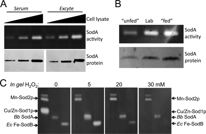
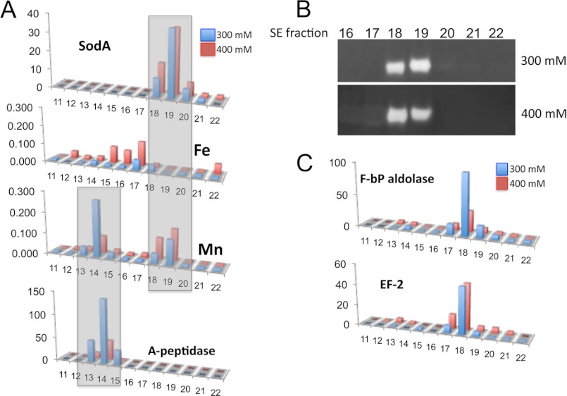
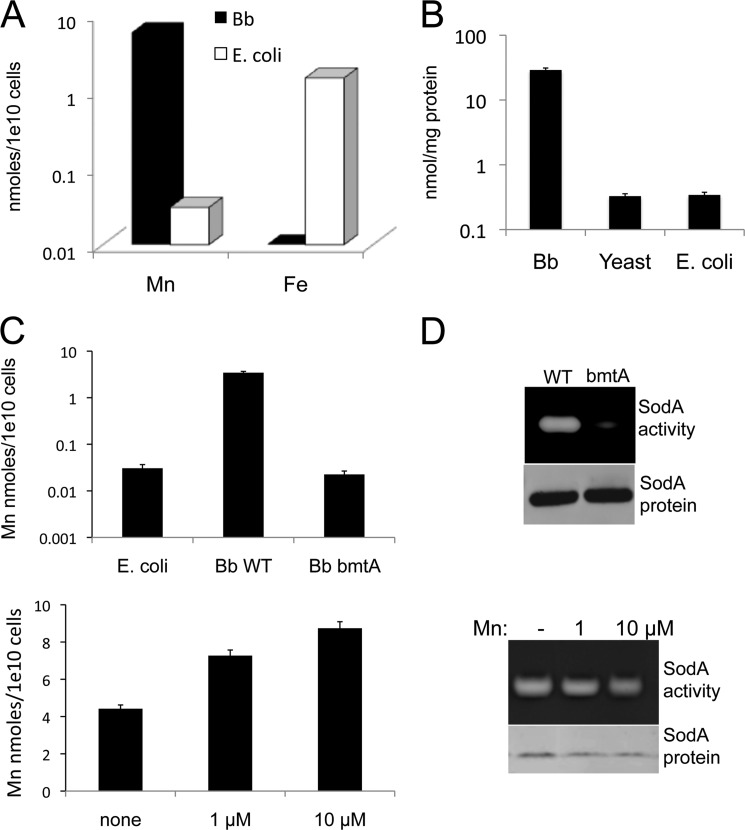
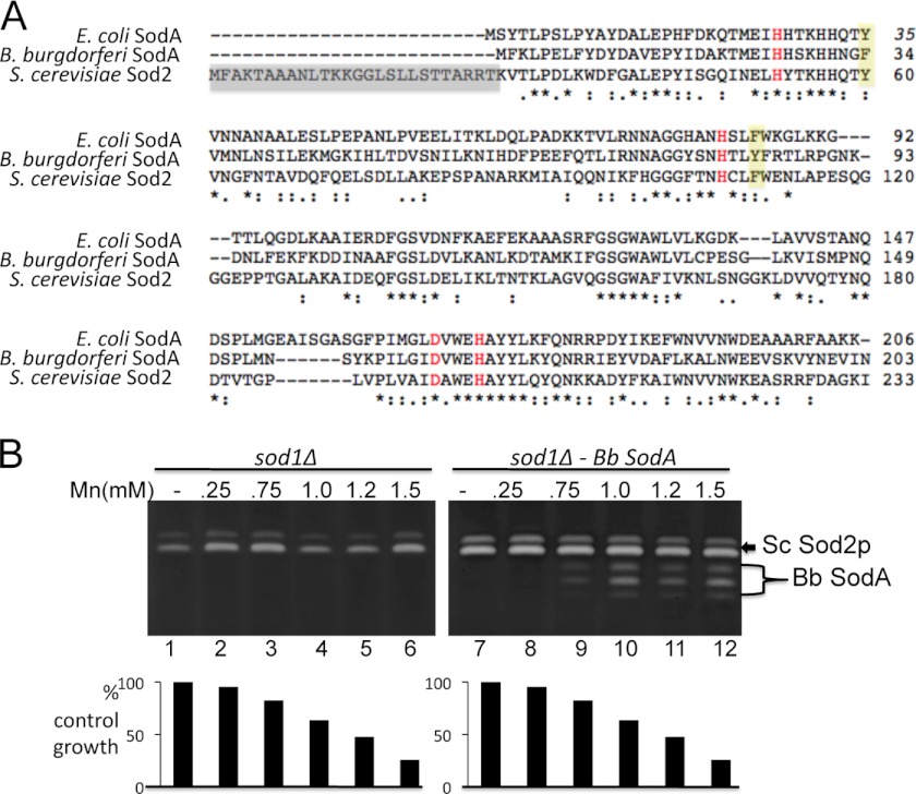
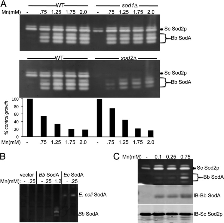
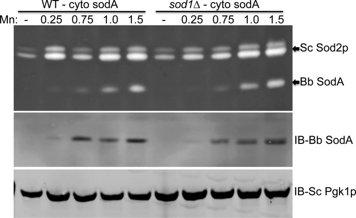
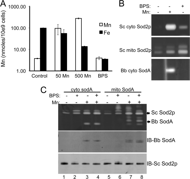
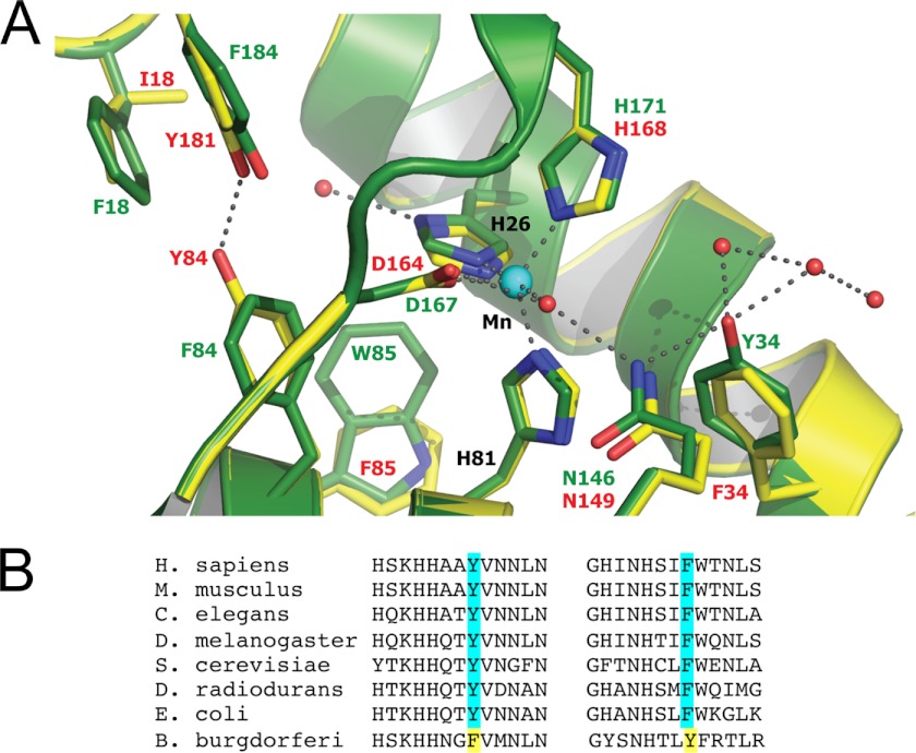
The Lyme disease pathogen Borrelia burgdorferi represents a novel organism in which to study metalloprotein biology in that this spirochete has uniquely evolved with no requirement for iron. Not only is iron low, but we show here that B. burgdorferi has the capacity to accumulate remarkably high levels of manganese. This high manganese is necessary to activate the SodA superoxide dismutase (SOD) essential for virulence. Using a metalloproteomic approach, we demonstrate that a bulk of B. burgdorferi SodA directly associates with manganese, and a smaller pool of inactive enzyme accumulates as apoprotein. Other metalloproteins may have similarly adapted to using manganese as co-factor, including the BB0366 aminopeptidase. Whereas B. burgdorferi SodA has evolved in a manganese-rich, iron-poor environment, the opposite is true for Mn-SODs of organisms such as Escherichia coli and bakers' yeast. These Mn-SODs still capture manganese in an iron-rich cell, and we tested whether the same is true for Borrelia SodA. When expressed in the iron-rich mitochondria of Saccharomyces cerevisiae, B. burgdorferi SodA was inactive. Activity was only possible when cells accumulated extremely high levels of manganese that exceeded cellular iron. Moreover, there was no evidence for iron inactivation of the SOD. B. burgdorferi SodA shows strong overall homology with other members of the Mn-SOD family, but computer-assisted modeling revealed some unusual features of the hydrogen bonding network near the enzyme's active site. The unique properties of B. burgdorferi SodA may represent adaptation to expression in the manganese-rich and iron-poor environment of the spirochete.


Figures

FIGURE 1.
FIGURE 1.
Activity of B. burgdorferi SodA in its native host and its sensitivity toward peroxide. A and B, whole cell B. burgdorferi lysates were prepared from strain ML23 and were analyzed for SOD activity by native gel electrophoresis and nitro blue tetrazolium staining (SodA activity) and for SodA protein levels by immunoblot (SodA protein), as described under “Experimental Procedures.” A, cells were grown in BSK medium supplemented with either 6% (v/v) rabbit serum or with the synthetic serum substitute Ex-cyte. Wedge represents increasing levels of lysate protein analyzed: 2.5, 5, and 10 μg for SodA activity and 0.5, 1, and 2.5 μg for immunoblot. B, cells were grown in serum containing BSK under the following conditions: unfed, pH 7.6, 23 °C to simulate unfed tick host (25); lab, pH 7.6, 34 °C standard laboratory culture conditions; fed, pH 6.7, 37 °C to simulate post-blood meal conditions in the tick host (25). C, samples containing the indicated SOD enzymes were subjected to native gel electrophoresis, and prior to staining with nitro blue tetrazolium for SOD activity, the gel was incubated with the indicated concentrations of H2O2 as described under “Experimental Procedures.” Mn-Sod2p and Cu/Zn-Sod1p, S. cerevisiae SOD enzymes present in 50 μg of total yeast lysate protein. Bb SodA, 5.0 μg of B. burgdorferi lysate protein; Ec Fe-SodB, 0.06 units of purified SodB enzyme from E. coli (Sigma).
FIGURE 2.
FIGURE 2.
Multidimensional chromatography of B. burgdorferi lysates. Soluble B. burgdorferi lysates were resolved by anion exchange, and the 300 and 400 mm NaCl elutions were resolved by size exclusion (SE) chromatography; shown are results from fractions 11–22 of increasing retention time on size exclusion. A, fractions were subjected to either metal analysis by ICP-MS (Fe and Mn), where y axis units represent relative abundance, or proteomic analysis by trypsin digestion, LC/MS, and MS/MS (SodA and A-peptidase), where y axis units represent spectral counts. Shaded boxes indicate manganese peak overlaps with SodA and with amino peptidase-1 (gene BB0366). B, fractions were analyzed for SodA activity by the native gel assay. C, proteomic analysis of fractions to illustrate that the manganese in fraction 19 shows poor correlation with a fructose bisphosphate aldolase (F-bP aldolase gene BB0445) and elongation factor EF-2 (EF-2 gene BB0540).
FIGURE 3.
FIGURE 3.
Metal analysis of B. burgdorferi versus iron-philic organisms and effects on SodA activity. A, ICP MS analysis of manganese and iron was carried out with whole cell B. burgdorferi strain ML23 versus E. coli cells grown in BSK medium, as described under “Experimental Procedures.” B, AAS measurements of manganese in soluble protein lysates from B. burgdorferi (Bb) strain ML23, E. coli, and S. cerevisiae as described under “Experimental Procedures.” C, AAS analysis of whole cell manganese in B. burgdorferi strain 297 and the corresponding bmtA mutant compared with E. coli (top) and B. burgdorferi strain ML23 grown in BSK supplemented with the indicated concentrations of MnCl2 (bottom). D, SodA activity and protein levels were examined as in Fig. 1A in B. burgdorferi strain 297 and the corresponding bmtA mutant (top) and B. burgdorferi strain ML23 grown with the indicated concentrations of MnCl2 (bottom).
FIGURE 4.
FIGURE 4.
Targeting B. burgdorferi SodA to the mitochondria of S. cerevisiae. A, alignment of B. burgdorferi SodA with the Mn-Sod2p of S. cerevisiae and the Mn-SodA of E. coli using CLC sequencer viewer version 6.4 software. Asterisks mark identity, and dots represent similar residues; red marks, metal binding residues. The yellow shaded area marks the unique Phe-34 and Tyr-84 of B. burgdorferi SodA (see Fig. 8), and the gray shaded area shows the MLS of yeast Sod2p that was fused onto the bacterial SodA genes. B, a sod1Δ yeast strain was transformed where indicated with the pDA002 plasmid for expressing mitochondrial targeted B. burgdorferi SodA (lanes 7–12) and was grown with the indicated concentrations of MnCl2. Top, SOD activity was monitored by the native gel assay. The position of endogenous yeast Sod2p (Sc Sod2p) and heterologous B. burgdorferi SodA (Bb SodA) are indicated. Mitochondrial Sod2p runs as a doublet or triplet (14), and the same is true for B. burgdorferi SodA expressed in yeast mitochondria. Bottom, cell growth was monitored turbidimetrically at A600 nm and plotted as a percentage of control growth obtained in the absence of manganese.
FIGURE 5.
FIGURE 5.
B. burgdorferi SodA requires high manganese for activity and expression in yeast mitochondria. A, the indicated WT or sod mutants of S. cerevisiae expressing mitochondrial targeted B. burgdorferi SodA were tested for manganese activation of SOD activity and for manganese effects on cell growth (for WT and sod2Δ, bottom) as described in the legend to Fig. 4, except yeast Cu/Zn-Sod1p was inactivated by in-gel treatment with 5 mm H2O2. The positions of endogenous yeast Sod2p (Sc Sod2p) and heterologous B. burgdorferi SodA (Bb SodA) on the native activity gels are indicated. B, the sod1Δ sod2Δ expressing either empty vector pRS315 (58) or the mitochondrial targeted SodA from either B. burgdorferi or E. coli on plasmids pDA002 and pAN002 (14) was grown with the indicated concentrations of MnCl2 and analyzed for SOD activity by the native gel assay. The positions of mitochondrial targeted E. coli and B. burgdorferi SodA are indicated. C, WT yeast strains expressing mitochondrial B. burgdorferi SodA were grown in the presence of the indicated concentrations of MnCl2 and were subjected to native gel assays for SOD activity as in A (top) or to immunoblot (IB) analysis of B. burgdorferi SodA and yeast Sod2p protein levels (middle and bottom).
FIGURE 6.
FIGURE 6.
Expression of B. burgdorferi SodA in yeast cytosol. The WT yeast strain or sod1Δ mutant was transformed with plasmid pSA002 for expressing B. burgdorferi SodA in yeast cytosol, and cells were cultured with the indicated millimolar concentrations of MnCl2. Top, SOD activity was assayed as in Fig. 5C. Middle and bottom, immunoblot (IB) analysis was conducted as in Fig. 5C using antibodies directed against B. burgdorferi (Bb) SodA and yeast cytosolic (Sc) Pgk1p.
FIGURE 7.
FIGURE 7.
Iron chelation does not help activate B. burgdorferi SodA expressed in yeast. A, ICP-MS analysis of iron and manganese in whole cells of S. cerevisiae (Sc) grown in the presence of the indicated concentrations of MnCl2 or 100 μm BPS as described under “Experimental Procedures.” B, yeast strains transformed with pEL124 (37) for expressing yeast Sod2p in the cytosol (top) or with pDA002 for expressing mitochondrial B. burgdorferi (Bb) SodA (bottom) were grown in the presence of 1.0 mm MnCl2 (Mn: +) and/or 0.1 mm of the iron chelator BPS (BPS: +) and subjected to SOD activity analysis. C, yeast cells expressing either mitochondrial or cytosolic B. burgdorferi SodA were treated with manganese or BPS and tested for SOD activity as in B (top) and for levels of B. burgdorferi SodA and S. cerevisiae Sod2p by immunoblot as in Fig. 5C (middle and bottom). Error bars, standard deviation.
FIGURE 8.
FIGURE 8.
The predicted active site of B. burgdorferi SodA. A, a model of B. burgdorferi SodA was generated with the program MODELLER (53) using the 0.9 Å structure of E. coli SodA (Protein Data Bank accession number 1IX9) as the structural template. Residues of B. burgdorferi SodA are shown in yellow with numbering in red, and the equivalent positions in E. coli SodA are marked in green. Red balls indicate water molecules, and dotted lines represent hydrogen bonds or the coordination of the manganese ion (aqua ball) to its four amino acid ligands and a single water molecule. B, a comparison of Tyr-34 and Phe-84 in B. burgdorferi SodA with the equivalent positions in Mn-SOD molecules from the indicated organisms.

References

    1. Aguirre J. D., Culotta V. C. (2012) Battles with iron. Manganese in oxidative stress protection. J. Biol. Chem. 287, 13541–13548 - PMC - PubMed
    1. Wintjens R., Noël C., May A. C., Gerbod D., Dufernez F., Capron M., Viscogliosi E., Rooman M. (2004) Specificity and phenetic relationships of iron- and manganese-containing superoxide dismutases on the basis of structure and sequence comparisons. J. Biol. Chem. 279, 9248–9254 - PubMed
    1. Mizuno K., Whittaker M. M., Bächinger H. P., Whittaker J. W. (2004) Calorimetric studies on the tight-binding metal interactions of Escherichia coli manganese superoxide dismutase. J. Biol. Chem. 279, 27339–27344 - PubMed
    1. Iranzo O. (2011) Manganese complexes displaying superoxide dismutase activity. A balance between different factors. Bioorg. Chem. 39, 73–87 - PubMed
    1. Kang Y., He Y. X., Zhao M. X., Li W. F. (2011) Structures of native and Fe-substituted SOD2 from Saccharomyces cerevisiae. Acta Crystallogr. Sect. F Struct. Biol. Cryst. Commun. 67, 1173–1178 - PMC - PubMed

Publication types

MeSH terms