. 2013 Jun 10;333(2):205-12.
doi: 10.1016/j.canlet.2013.01.037. Epub 2013 Jan 29.

Generation of erythroid cells from fibroblasts and cancer cells in vitro and in vivo

Affiliations

Generation of erythroid cells from fibroblasts and cancer cells in vitro and in vivo

Shiwu Zhang et al. Cancer Lett. .

Abstract

Bone marrow is generally considered the main source of erythroid cells. Here we report that a single hypoxia-mimic chemical, CoCl2, can increase the size of fibroblasts and cancer cells and lead to formation of polyploidy giant cells (PGCs) or polyploidy giant cancer cells (PGCCs), activation of stem cell marker expression, increased growth of normal and cancer spheroid, and lead to differentiation of the fibroblasts and epithelial cells toward erythroid lineage expressing hemoglobins both in vitro and in vivo. Immunohistochemical examination demonstrated that these cells are predominantly made of embryonic hemoglobins, with various levels of fetal and adult hemoglobins. Ectopic expression of c-Myc induced the generation of nucleated erythoid cells expressing variable levels of embryonic and fetal hemoglobins. Generation of these erythroid cells can be also observed via histological examination of other cancer cell lines and human tumor samples. These data suggest that normal and solid cancer cells can directly generate erythroid cells to obtain oxygen in response to hypoxia and may explain the ineffectiveness of conventional anti-angiogenic therapies for cancer, which are directed at endothelium-dependent vessels, and offer new targets for intervention.


Figures

Fig. 1
Fig. 1
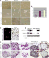
Generation of spheroids and erythroid cells after CoCl2 in fibroblast lines. (A) CoCl2-induced formation of spheroids in fibroblast lines. The morphology of NOF151 (a), NOF151hT (b), and NOF151p53ihT (c) cells is shown (×10). After CoCl2 treatment, the regularly sized cells were killed, whereas a few PGCs of NOF151 (d), NOF151hT (e), and NOF151p53ihT (f) survived. These PGCs can form spheroids, which were shown to be NOF151 (g), NOF151hT (h), and NOF151p53ihT (i) (×10). (B) Comparison of the numbers of spheroids in NOF151, NOF151hT, and NOF151p53ihT cells. (C) SOX-2 staining of NOF151hT and NOFp53ihT spheroids. (a and c) NOF151hT and NOFp53ihT spheroids were positive for SOX-2 immunofluorescent stain (×20) (b and d) NOF151hT and NOFp53ihT spheroids were positive for SOX-2 immunochemical stain (×20). (D) Erythroid cell differentiation of NOF151hT and NOF151p53ihT cells induced by CoCl2 treatment in vitro. H&E staining of NOF151hT and NOFp53ihT spheroid slides showed that erythroid cells generated in NOF151hT (a–c) and NOF151p53ihT (f–h) after the treatment (H&E staining, ×20); (b) and (g) are the high-power views of (a) and (f), respectively, to clearly show the erythroid cells budding from the spheroids (black arrowheads) (H&E staining, ×20). Spheroids were positive for hemoglobin-β/γ/δ/ε (d and i) and hemoglobin- δ (e and j) (immunohistochemical staining, ×20). (E) Western blot analysis of hemoglobin expression in NOF151hT and NOF151p53ihT cells with and without CoCl2 treatment.
Fig. 2
Fig. 2
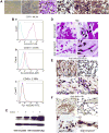
Erythroid cells differentiation in NOF137p53ihTc-Myc and xenografted tissue. (A) Erythroid cells differentiation of NOF137p53ihTc-Myc treated with CoCl2. (a) Control NOF137p53ihT (×10). (b) Suspension cells generated by NOF137p53ihTc-Myc after CoCl2 treatment (×10). (c)H&E staining of paraffin-embedded suspension cells (×20). (d–f) Suspension cells were positive for hemoglobin-β/γ/δ/ε(d), fetal (e), and -δ (f) (×20). (B) Flow cytometric analysis of suspension cells with CD71 (a), CD34 (b), and CD45 (c) after CoCl2 treatment. The blue lines show PI staining only and the red lines are for PI-negative cells and CD71-positive (a), PI-negative cells and CD34-positive (b), and PI-negative cells and CD45-positive (c). (C) Western blot results of c-Myc expression. (D) Erythroid cell differentiation in xenografted CoCl2-treated NOF151p53ihT and NOF137p53ihT spheroids (H&E staining, ×20). Erythroid cells generated from NOF151p53ihT (a and b) and NOF137p53ihT (c and d) mouse xenografts (black arrowheads) (H&E staining, ×20). (E) Immunohistochemical staining of mouse xenografts with human specific anti-mitochondrial antibodies. (a and b) Anti-mitochondrial antibody showed specific staining against mitochondrial in human tissue (a) and no staining in mouse tissue (b)(×20). (c and d) Human origin of the cells in mouse xenografts formed by NOF151p53ihT (c) and NOF137p53ihT (d) was confirmed using immunohistochemical staining with anti-mitochondrial antibodies (black arrowheads, ×20). (F) Immunohistochemical staining with human specific anti-hemoglobin-δ antibody. The specificity of monoclonal antibody against human hemoglobin- δ was shown by its interaction with the erythroid cells of human origin (a) but not with erythroid cells of mouse origin (b). The erythroid cells generated by NOF151p53ihT (c) and NOF137p53ihT (d) were positive for anti-hemoglobin- δ antibodies (black arrowheads, ×20). (For interpretation of the references to colour in this figure legend, the reader is referred to the web version of this article.)
Fig. 3
Fig. 3
Generation of erythroid cells in cancer cell lines. (A) Formation of BT-549 spheroids after CoCl2 treatment. (a) Control BT-549 without CoCl2 treatment (×10). (b) PGCCs of BT-549 (large arrowheads) survived CoCl2 treatment and produced regularly sized cells (small arrowheads) (×10). (c) BT-549 spheroid formation after CoCl2 treatment (×10). (d and e) Immunohistochemical stain for CD44 (c) and CD133 (d) (×20). (f) Immunofluorescent stain for SOX-2 (×20). (g) Immunohistochemical stain of spheroids for SOX-2 (×20). (B) Generation of erythroid cells by BT-549 cells in vitro. (a) H&E stain of spheroids showing erythroid cells generated by BT-549 cells (×20). (b) High–power view of erythroid cells (H&E stain, ×40). (c) Erythroid cells budding from cancer cells (black arrow points; HE staining, ×20). (d–h) Immunohistochemical staining of erythroid cells for hemoglobin-α,-β/γ/ε/δ, -ε, fetal hemoglobin, and hemoglobin-ζ (×20). (C) Erythroid cells differentiation in mouse xenografted tumor from CoCl2-treated BT-549 injection. (a–c) H&E staining of tumor tissue. (a) Many erythroid cells around tumor cells. (b) Erythroid cells appearing in the cytoplasm of tumor cells. (c) Erythroid cells budding from tumor cells (black arrowheads) (×20). (d) Immunohistochemical staining with human specific anti-hemoglobin- δ antibody confirming the human origin of erythroid cells (×20).
Fig. 4
Fig. 4
Generation of erythroid cells and hemoglobin in cancer cell lines and tissue. (A) Generation of erythroid cells by cancer cell lines. Erythroid cells can be identified from spheroids generated by, MDA-MB-231 (a), FTE187SV40hT-Hras (b), and Phoenix (c) cells in vitro after CoCl2 treatment (black arrowheads, ×20). (B) Generation of erythroid cells and hemoglobin by human ovarian cancer cells. (a) Smear of cancer cells in the medium confirming the generation of erythroid cells by high-grade ovarian cancer cells in vitro (black arrowheads, ×20). (b) Erythroid cells in the cytoplasm of high-grade ovarian carcinoma cells (×20). (c and d) Numerous round and red hemoglobin bodies surrounding and within human ovarian cancer cells. (e–h) Staining of round and red hemoglobin bodies for hemoglobin- α (e); hemoglobin-β/γ/ε/δ (f); fetal hemoglobin (g); and hemoglobin-ζ (h) in the same fields shown in c (black arrowheads, ×20). (For interpretation of the references to colour in this figure legend, the reader is referred to the web version of this article.)

References

    1. Palis J, Segel GB. Developmental biology of erythropoiesis. Blood Rev. 1998;12:106–114. - PubMed
    1. Fried W. Erythropoietin and erythropoiesis. Exp Hematol. 2009;37:1007–1015. - PubMed
    1. Tsuchida E, Komatsu T, Hamamatsu K, Matsukawa Y, Tajima A, Yoshizu A, Izumi Y, Kobayashi K. Exchange transfusion with albumin-heme as an artificialO2-infusion into anesthetized rats: physiological responses, O2-delivery, and reduction of the oxidized hemin sites by red blood cells. Bioconjug Chem. 2000;11:46–50. - PubMed
    1. Cohen WD, Bartelt D, Jaeger R, Langford G, Nemhauser I. The cytoskeletal system of nucleated erythrocytes. I. Composition and function of major elements. J Cell Biol. 1982;93:828. - PMC - PubMed
    1. Guo Y, Niu C, Breslin P, Tang M, Zhang S, Wei W, Kini AR, Paner GP, Alkan S, Morris SW, Diaz M, Stiff PJ, Zhang J. C-Myc-mediated control of cell fate in megakaryocyte-erythrocyte progenitors. Blood. 2009;114:2097–2106. - PMC - PubMed

Publication types

MeSH terms