

Distribution of secretagogin-containing neurons in the basal forebrain of mice, with special reference to the cholinergic corticopetal system
- PMID: 23376788
- PMCID: PMC3628291
- DOI: 10.1016/j.brainresbull.2013.01.009
Distribution of secretagogin-containing neurons in the basal forebrain of mice, with special reference to the cholinergic corticopetal system
Abstract
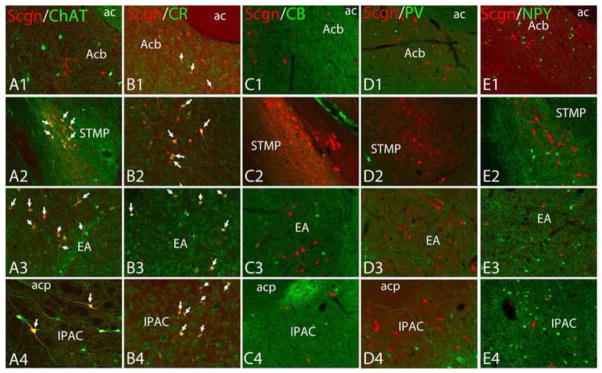
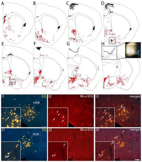
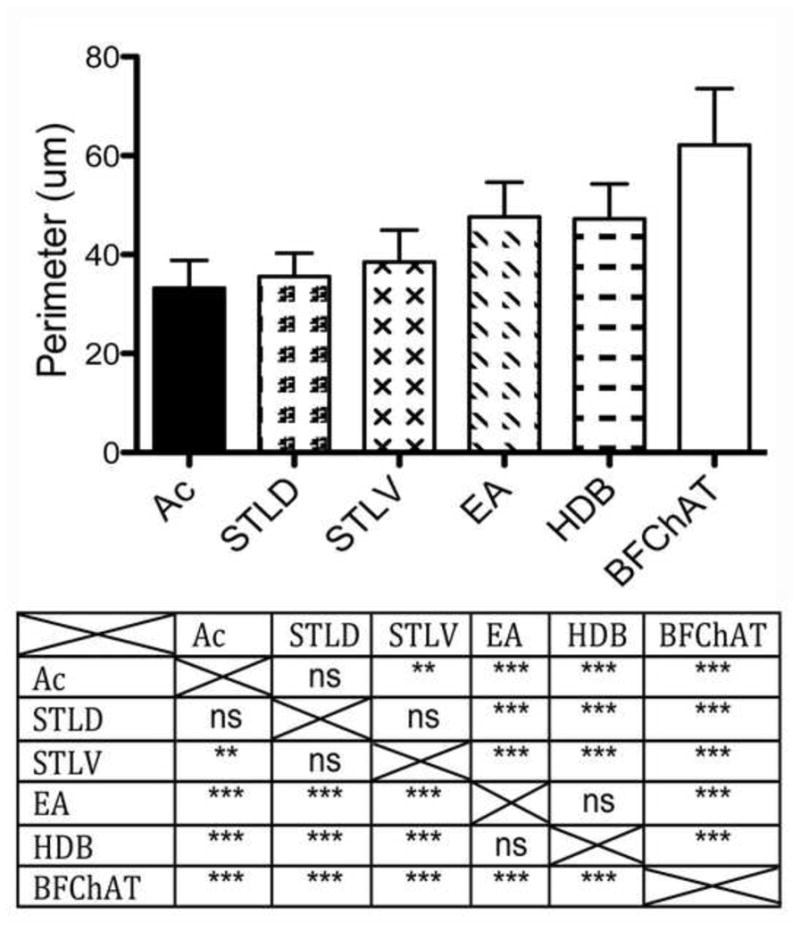
Cholinergic and GABAergic corticopetal neurons in the basal forebrain play important roles in cortical activation, sensory processing, and attention. Cholinergic neurons are intermingled with peptidergic, and various calcium binding protein-containing cells, however, the functional role of these neurons is not well understood. In this study we examined the expression pattern of secretagogin (Scgn), a newly described calcium-binding protein, in neurons of the basal forebrain. We also assessed some of the corticopetal projections of Scgn neurons and their co-localization with choline acetyltransferase (ChAT), neuropeptide-Y, and other calcium-binding proteins (i.e., calbindin, calretinin, and parvalbumin). Scgn is expressed in cell bodies of the medial and lateral septum, vertical and horizontal diagonal band nuclei, and of the extension of the amygdala but it is almost absent in the ventral pallidum. Scgn is co-localized with ChAT in neurons of the bed nucleus of the stria terminalis, extension of the amygdala, and interstitial nucleus of the posterior limb of the anterior commissure. Scgn was co-localized with calretinin in the accumbens nucleus, medial division of the bed nucleus of stria terminalis, the extension of the amygdala, and interstitial nucleus of the posterior limb of the anterior commissure. We have not found co-expression of Scgn with parvalbumin, calbindin, or neuropeptide-Y. Retrograde tracing studies using Fluoro Gold in combination with Scgn-specific immunohistochemistry revealed that Scgn neurons situated in the nucleus of the horizontal limb of the diagonal band project to retrosplenial and cingulate cortical areas.
Copyright © 2013 Elsevier Inc. All rights reserved.
Figures

Similar articles
-
Secretagogin is a Ca2+-binding protein identifying prospective extended amygdala neurons in the developing mammalian telencephalon.Eur J Neurosci. 2010 Jun;31(12):2166-77. doi: 10.1111/j.1460-9568.2010.07275.x. Epub 2010 Jun 7. Eur J Neurosci. 2010. PMID: 20529129 Free PMC article.
-
GABAergic and cholinergic basal forebrain and preoptic-anterior hypothalamic projections to the mediodorsal nucleus of the thalamus in the cat.Neuroscience. 1998 Jul;85(1):149-78. doi: 10.1016/s0306-4522(97)00573-3. Neuroscience. 1998. PMID: 9607710
-
Estrogen receptor immunoreactivity within subregions of the rat forebrain: neuronal distribution and association with perikarya containing choline acetyltransferase.Brain Res. 1999 Dec 4;849(1-2):253-74. doi: 10.1016/s0006-8993(99)01960-5. Brain Res. 1999. PMID: 10592312
-
Projections of GABAergic and cholinergic basal forebrain and GABAergic preoptic-anterior hypothalamic neurons to the posterior lateral hypothalamus of the rat.J Comp Neurol. 1994 Jan 8;339(2):251-68. doi: 10.1002/cne.903390206. J Comp Neurol. 1994. PMID: 8300907
-
Calcium-binding protein, secretagogin, specifies the microcellular tegmental nucleus and intermediate and ventral parts of the cuneiform nucleus of the mouse and rat.Neurosci Res. 2018 Sep;134:30-38. doi: 10.1016/j.neures.2018.01.005. Epub 2018 Feb 3. Neurosci Res. 2018. PMID: 29366872
Cited by
-
Impact of hypothalamic reactive oxygen species in the regulation of energy metabolism and food intake.Front Neurosci. 2015 Feb 24;9:56. doi: 10.3389/fnins.2015.00056. eCollection 2015. Front Neurosci. 2015. PMID: 25759638 Free PMC article. Review.
-
20 Years of Secretagogin: Exocytosis and Beyond.Front Mol Neurosci. 2019 Feb 12;12:29. doi: 10.3389/fnmol.2019.00029. eCollection 2019. Front Mol Neurosci. 2019. PMID: 30853888 Free PMC article.
-
Developmental and adult characterization of secretagogin expressing amacrine cells in zebrafish retina.PLoS One. 2017 Sep 26;12(9):e0185107. doi: 10.1371/journal.pone.0185107. eCollection 2017. PLoS One. 2017. PMID: 28949993 Free PMC article.
-
The cholinergic basal forebrain in the ferret and its inputs to the auditory cortex.Eur J Neurosci. 2014 Sep;40(6):2922-40. doi: 10.1111/ejn.12653. Epub 2014 Jun 19. Eur J Neurosci. 2014. PMID: 24945075 Free PMC article.
-
Basal Forebrain Projections to the Retrosplenial and Cingulate Cortex in Rats.J Comp Neurol. 2025 Feb;533(2):e70027. doi: 10.1002/cne.70027. J Comp Neurol. 2025. PMID: 39924777 Free PMC article.
References
-
- Attems J, Ittner A, Jellinger K, Nitsch RM, Maj M, Wagner L, Gotz J, Heikenwalder M. Reduced secretagogin expression in the hippocampus of P301L tau transgenic mice. J Neural Transm. 2011;118:737–745. - PubMed
-
- Attems J, Quass M, Gartner W, Nabokikh A, Wagner L, Steurer S, Arbes S, Lintner F, Jellinger K. Immunoreactivity of calcium binding protein secretagogin in the human hippocampus is restricted to pyramidal neurons. Exp Gerontol. 2007;42:215–222. - PubMed
-
- Bu J, Sathyendra V, Nagykery N, Geula C. Age-related changes in calbindin-D28k, calretinin, and parvalbumin-immunoreactive neurons in the human cerebral cortex. Exp Neurol. 2003;182:220–231. - PubMed
Publication types
MeSH terms
Substances
Grants and funding
LinkOut - more resources
Full Text Sources
Other Literature Sources
