. 2013 Apr 1;41(6):3734-47.
doi: 10.1093/nar/gkt047. Epub 2013 Feb 1.

Transcribed pseudogene ψPPM1K generates endogenous siRNA to suppress oncogenic cell growth in hepatocellular carcinoma

Affiliations

Transcribed pseudogene ψPPM1K generates endogenous siRNA to suppress oncogenic cell growth in hepatocellular carcinoma

Wen-Ling Chan et al. Nucleic Acids Res. .

Abstract

Pseudogenes, especially those that are transcribed, may not be mere genomic fossils, but their biological significance remains unclear. Postulating that in the human genome, as in animal models, pseudogenes may function as gene regulators through generation of endo-siRNAs (esiRNAs), antisense RNAs or RNA decoys, we performed bioinformatic and subsequent experimental tests to explore esiRNA-mediated mechanisms of pseudogene involvement in oncogenesis. A genome-wide survey revealed a partial retrotranscript pseudogene ψPPM1K containing inverted repeats capable of folding into hairpin structures that can be processed into two esiRNAs; these esiRNAs potentially target many cellular genes, including NEK8. In 41 paired surgical specimens, we found significantly reduced expression of two predicted ψPPM1K-specific esiRNAs, and the cognate gene PPM1K, in hepatocellular carcinoma compared with matched non-tumour tissues, whereas the expression of target gene NEK8 was increased in tumours. Additionally, NEK8 and PPM1K were downregulated in stably transfected ψPPM1K-overexpressing cells, but not in cells transfected with an esiRNA1-deletion mutant of ψPPM1K. Furthermore, expression of NEK8 in ψPPM1K-transfected cells demonstrated that NEK8 can counteract the growth inhibitory effects of ψPPM1K. These findings indicate that a transcribed pseudogene can exert tumour-suppressor activity independent of its parental gene by generation of esiRNAs that regulate human cell growth.


Figures

Figure 1.
Figure 1.
Workflow for identification of pseudogene-derived esiRNA–target interactions. Using a systematic computational procedure of homologous sequence alignment between a collection of transcribed pseudogenes and known functional sRNAs, we identified pseudogene-derived esiRNAs and verified these by reference to available Illumina-Solexa reads, and subsequently by reference to regulated protein-coding target genes (see ‘Materials and Methods’ section).
Figure 2.
Figure 2.
Schematic representation of ψPPM1K and its parental gene, PPM1K. (a) ψPPM1K is located on chromosome 4 proximal to PPM1K. (b) Alignment of ψPPM1K and its cognate gene PPM1K indicates that ψPPM1K is a reversed-transcribed partial antisense copy of its parental gene, PPM1K, with >79% sequence homology in the 155–456-nt region.
Figure 3.
Figure 3.
Candidate ψPPM1K-derived esiRNAs and their targets. (a) Location and read counts of transcribed ψPPM1K RNA from sRNA deep sequencing data. The candidate precursor esiRNAs derived from the ψPPM1K transcript are shown by green (24–144 nt) and red rectangles (170–273 nt) with read counts 4 and 32, respectively. The other putative esiRNA in the 328–455-nt region (pink rectangle) with 39 read counts was excluded from further analysis, as it is located within the protein-coding gene region that shows a high degree of similarity between ψPPM1K and PPM1K. (b) Sequences of precursor esiRNAs, with esiRNAs shown in colour. (c) The alignment of esiRNA3 mapping to PPM1K gene. (d) Mfold was used to predict the hairpin structure of precursor esiRNA1 with MFE −40.4. The predicted mature esiRNA1 sequence is depicted in green. (e) Matches of esiRNA1 and esiRNA2 sequences with target gene NEK8 and parental gene PPM1K. Canonical pairings, solid lines; non-canonical pairings (G:U), dotted lines. (f) Expression profiles of ψPPM1K, PPM1K and NEK8 in HCC tissues/cells (GSE6222).
Figure 4.
Figure 4.
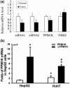
Expression patterns of HCC tissues and cell lines. (a) RT–qPCR results showing relative expression levels of two esiRNA precursors, PPM1K and NEK8, in HCC tumour and paired non-tumour tissues from 41 HCC patients (*P ≤ 0.01). (b) RNA levels of PPM1K and ψPPM1K in HepG2 and Huh-7 cells. RT–qPCR results show that PPMIK expression was higher in Huh7 than in HepG2 cells. The expression of ψPPM1K was higher than PPMIK in both cell lines (n = 7). *P < 0.05 compared with PPM1K in HepG2; #P < 0.05 compared with PPM1K in Huh-7.
Figure 5.
Figure 5.
Effect of overexpressed ψPPM1K on cell growth and clonogenic activity in transfected Huh-7 cell clones. (a) HCC line Huh-7 and HepG2 cells were transfected with ψPPM1K-expressing recombinant plasmid to isolate stably transfected cell clones (TPG1, TPG2 and TPG7 for Huh-7; TPG for HepG2) by G418 selection, or with empty vector plasmid for G418-selected control mock2 cells (Vector). Equivalent amounts of total RNA from each cell clone were used for RT–PCR/gel electrophoresis analysis of ψPPM1K and GAPDH mRNA. (b) All three ψPPM1K-expressing cell lines have a slower proliferation rate than the vector control cell line (TPG7 cells: P = 0.036 for Day 2, P = 0.018 for Day 4; TPG1 cells: P = 0.045 for Day 4). (c) Serial photographs of the same colonies at Day 5, Day 7 and Day 9 showing the 2D growth of mock2, TPG1, TPG2 and TPG7 transfected Huh-7 clones on plastic culture dishes. (d) Clonogenic activity of mock2, TPG1, TPG2 and TPG7 was determined in soft-agar cultures incubated under normoxic 19% O2 and hypoxic 3% O2 conditions in 5% CO2 incubators for 12 days. Numbers of colonies formed per 1000 cells were counted for mock2, TPG1, TPG2 and TPG7 cell clones. Decreased clonogenicity compared with the mock2 control was calculated for TPG1 (P = 1.54E-06), for TPG2 (P = 0.0001) and for TPG7 (P = 0.013) in normoxic conditions; for TPG1 (P = 2.22E-05), for TPG2 (P = 5.71E-05) and for TPG7 (P = 0.0011) in hypoxic conditions. (*P < 0.05).
Figure 6.
Figure 6.
Expression of predicted target genes in Huh-7 and HepG2 HCC cell clones transfected with ψPPM1K. (a) The mRNA levels of three potential esiRNA1-targeted genes (NEK8, TBRG1 and BMPR2) were analysed by RT–qPCR. NEK8 was downregulated in all three ψPPM1K-expressing Huh-7 cell lines relative to the vector control cells (*P = 0.033 in TPG7 cells). (b) NEK8 mRNA levels were reduced in the ψPPM1K-expressing HepG2-TPG cell line relative to the vector control cells (*P = 0.047).
Figure 7.
Figure 7.
Expression of ψPPM1K-dervied esiRNAs. (a) ψPPM1K-dervied esiRNAs detection by northern blots. Total RNA from human hepatoma cell Huh-7 was resolved by agarose electrophoresis and then transferred to an NC membrane. The NC membrane was hybridized with a biotinylated esiRNA probe overnight, washed and incubated with horseradish peroxidase-conjugated avidin. Finally, esiRNAs signals were detected by chemiluminescence. (b) Expression of esiRNA1 in the ψPPM1K-expressing Huh-7 stable cell line (TPG7) and the control cell line (Vector). The esiRNA1 levels were determined using a TaqMan MicroRNA Assay (Applied Biosystems) and were significantly higher in TPG7 cells than in vector control cells (*P < 0.05).
Figure 8.
Figure 8.
Expression of NEK8 in Huh-7 cells transfected with synthetic siRNA1. (a) The sequence of synthetic siRNA1. (b) Huh-7 cells were transfected with synthetic siRNA1 or with negative control siRNA (NC) for 48 h. Total RNA was isolated, and the NEK8 mRNA level was analysed by RT–qPCR. NEK8 was downregulated in the Huh-7 cells transfected with the synthetic siRNA1 relative to those transfected with negative control siRNA (*P = 0.001), implying that synthetic siRNA homologous to esiRNA1 can directly downregulate NEK8 gene expression in Huh-7 cells. (c) Expression of NEK8 in an esiRNA1-deletion mutant cell line. NEK8 mRNA levels in the ψPPM1K-expressing HepG2 cell line (TPG), the esiRNA1-deleted ψPPM1K-expressing cell line (mTPG) and the vector control cell line (Vector) were analysed by RT–qPCR. The results showed that NEK8 was downregulated in TPG cells (*P = 0.025 relative to vector control cells), but not in mTPG cells (P = 0.534 relative to vector control cells). (d) Growth of Huh-7 TPG7 cells transfected with either NEK8-overexpressing plasmid or empty vector analysed by cell proliferation assay. The proliferation rate of NEK8-overexpressing TPG7 cells was significantly higher than vector-transfected control cells (*P = 0.041 at Day 4).
Figure 9.
Figure 9.
ψPPM1K alters PPM1K expression and mitochondrial function. (a) ψPPM1K-derived esiRNA1 downregulates PPM1K expression. PPM1K mRNA levels in the ψPPM1K-expressing HepG2 cell line (TPG), the esiRNA1-deleted ψPPM1K-expressing cell line (mTPG) and the vector control cell line (Vector) were analysed by RT–qPCR. The results showed that PPM1K was downregulated in TPG cells (*P = 0.018 relative to vector control cells), but not in mTPG cells. (b) Fluorescence-activated cell sorting analysis shows no significant differences of mitochondrial Rh123 uptake by the four transfected Huh-7 clones. (*P < 0.01). (c) Release of Rh123 from mitochondria (0.5–18 h) was significantly faster in ψPPM1K-expressing Huh-7 cell line (TPG7) cells (84.59%) than Vector control cells (81.32%) (P = 0.031 for 0.5 h; P = 1.3E-06 for 18 h).
Figure 10.
Figure 10.
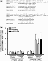
miRNA regulation of PPM1K and ψPPM1K. (a) Alignments of miRNA sequences with PPM1K and ψPPM1K. (b) Expression of PPM1K and ψPPM1K in miRNA-transfected TPG7 and mock cells. PPM1K is significantly downregulated in TPG7 and mock cells, and ψPPM1K is downregulated in mock cells, relative to a stable negative control (siCon) after miR-3174 transfection for 48 h (*P ≤ 0.01).
Figure 11.
Figure 11.
Possible genetic regulatory mechanisms involving ψPPM1K. Transcripts of ψPPM1K are exported to the cytoplasm where dsRNAs are cut from hairpin structures by Dicer into esiRNAs, which then interact with protein-coding target genes. Our results indicate that ψPPM1K-derived esiRNA1 may inhibit HCC cell proliferation through downregulation of NEK8, as well as by decreasing expression of PPM1K and alteration of mitochondrial activation.

Similar articles

Cited by

References

    1. Torrents D, Suyama M, Zdobnov E, Bork P. A genome-wide survey of human pseudogenes. Genome Res. 2003;13:2559–2567. - PMC - PubMed
    1. Mighell AJ, Smith NR, Robinson PA, Markham AF. Vertebrate pseudogenes. FEBS Lett. 2000;468:109–114. - PubMed
    1. Balasubramanian S, Zheng D, Liu YJ, Fang G, Frankish A, Carriero N, Robilotto R, Cayting P, Gerstein M. Comparative analysis of processed ribosomal protein pseudogenes in four mammalian genomes. Genome Biol. 2009;10:R2. - PMC - PubMed
    1. Harrison P, Yu Z. Frame disruptions in human mRNA transcripts, and their relationship with splicing and protein structures. BMC Genomics. 2007;8:371. - PMC - PubMed
    1. Harrison PM, Zheng D, Zhang Z, Carriero N, Gerstein M. Transcribed processed pseudogenes in the human genome: an intermediate form of expressed retrosequence lacking protein-coding ability. Nucleic Acids Res. 2005;33:2374–2383. - PMC - PubMed

Publication types

MeSH terms