Review
. 2013 Oct;70(19):3571-89.
doi: 10.1007/s00018-013-1261-0. Epub 2013 Feb 3.

O death where is thy sting? Immunologic tolerance to apoptotic self

Affiliations
Review

O death where is thy sting? Immunologic tolerance to apoptotic self

Buvana Ravishankar et al. Cell Mol Life Sci. 2013 Oct.

Abstract

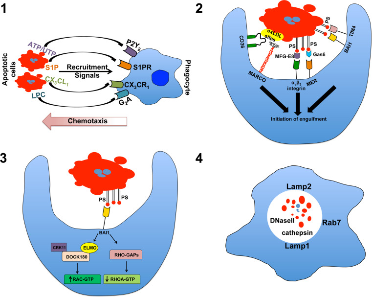
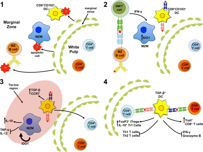
In higher organisms, innate scavenging cells maintain physiologic homeostasis by removal of the billions of apoptotic cells generated on a daily basis. Apoptotic cell removal requires efficient recognition and uptake by professional and non-professional phagocytic cells, which are governed by an array of soluble and apoptotic cell-integral signals resulting in immunologically silent clearance. While apoptosis is associated with profound suppression of adaptive and innate inflammatory immunity, we have only begun to scratch the surface in understanding how immunologic tolerance to apoptotic self manifest at either the molecular or cellular level. In the last 10 years, data has emerged implicating professional phagocytes, most notably stromal macrophages and CD8α(+)CD103(+) dendritic cells, as critical in initiation of the regulatory cascade that will ultimately lead to long-term whole-animal immune tolerance. Importantly, recent work by our lab and others has shown that alterations in apoptotic cell perception by the innate immune system either by removal of critical phagocytic sentinels in secondary lymphoid organs or blockage of immunosuppressive pathways leads to pronounced inflammation with a breakdown of tolerance towards self. This challenges the paradigm that apoptotic cells are inherently immunosuppressive, suggesting that apoptotic cell tolerance is a "context-dependent" event.


Figures

Fig. 1
Fig. 1
The four steps in efferocytosis. 1 In the “find me” stage, cellular commitment to apoptosis results in the production/release of chemotactic agents that recruit and, perhaps, activate local phagocytes priming them for phagocytic uptake of the cellular corpse. 2 During the “tickle me” stage, phagocytes encountering dying cells interact by a variety of receptors that can directly or indirectly (via bridging molecules) recognize exposure of native and modified forms of the membrane-integral lipid phosphatidylserine (PS) or other exposed, modified self-constitutions (the example shown here is MARCO and CD36-binding modified lipoproteins). 3 During the “eat me” stage, proper ligand/receptor engagement activates downstream signal transduction activating ELMO and Rac-dependent cytoskeletal changes resulting in plasma membrane invagination and internalization of the apoptotic cell. 4 During the “process me” phase, the apoptotic cell is degraded in Lamp+ lysosomes allowing for repurposing of the apoptotic material for antigen presentation and elicitation of adaptive, tolerogenic immunity. oxLDL oxidized low-density lipoprotein
Fig. 2
Fig. 2
Hypothetical model of induced splenic tolerance to apoptotic cells. 1 Circulating apoptotic cells enter the spleen through the marginal sinus traveling towards the red pulp. The apoptotic cells are captured in the marginal zone (MZ), primarily by marginal zone macrophages (MARCO+ MZM) and resident CD8+ DCs. However, MZ B cells also capture apoptotic cells in a B cell receptor-dependent manner. 2 Activated MZ B cells present either endogenous lipids or lipids derived from captured, internalized apoptotic cells in CD1d molecules to invariant NKT cells stimulating rapid production of IFN-γ. IFN-γ stimulates the MZMs to induce intracellular expression of the immunoregulatory enzyme IDO1. 3 IDO1 acts on MZMs in an autocrine and paracrine fashion promoting apoptotic cell-induced IL-10 production while inhibiting proinflammatory cytokine synthesis. Moreover, induction of IDO creates a zone of low tryptophan promoting stress response driven TGF-β production in CD8+ CD103+ tolerogenic DCs. The tolerogenic DCs also up-regulate surface expression of the chemokine receptor CCR7 promoting migration into the PALS. 4 Once in the PALS the TGF-β+CD8+ CD103+ DCs present apoptotic cell antigens by both MHC I and MHC II-dependent mechanisms promoting development of FoxP3+ Tregs, IL-10+ Tr1 T cells (i.e., IL-10+ CD4+ T cells), and Trail+ CD8+ T cells while inhibiting inflammatory CD4+ and CD8+ T cell function, promoting T cell anergy and deletion

Similar articles

Cited by

References

    1. Kerr JF, Wyllie AH, Currie AR. Apoptosis: a basic biological phenomenon with wide-ranging implications in tissue kinetics. Br J Cancer. 1972;26(4):239–257. doi: 10.1038/bjc.1972.33. - DOI - PMC - PubMed
    1. Elliott MR, Ravichandran KS. Clearance of apoptotic cells: implications in health and disease. J Cell Biol. 2010;189(7):1059–1070. doi: 10.1083/jcb.201004096. - DOI - PMC - PubMed
    1. Platt N, Suzuki H, Kodama T, Gordon S. Apoptotic thymocyte clearance in scavenger receptor class A-deficient mice is apparently normal. J Immunol. 2000;164(9):4861–4867. - PubMed
    1. Clancy RM, Neufing PJ, Zheng P, O’Mahony M, Nimmerjahn F, Gordon TP, Buyon JP. Impaired clearance of apoptotic cardiocytes is linked to anti-SSA/Ro and -SSB/La antibodies in the pathogenesis of congenital heart block. J Clin Invest. 2006;116(9):2413–2422. - PMC - PubMed
    1. Albert ML, Pearce SF, Francisco LM, Sauter B, Roy P, Silverstein RL, Bhardwaj N. Immature dendritic cells phagocytose apoptotic cells via alphavbeta5 and CD36, and cross-present antigens to cytotoxic T lymphocytes. J Exp Med. 1998;188(7):1359–1368. doi: 10.1084/jem.188.7.1359. - DOI - PMC - PubMed