

Utility of multiparametric 3-T MRI for glioma characterization
- PMID: 23377234
- PMCID: PMC4209475
- DOI: 10.1007/s00234-013-1145-x
Utility of multiparametric 3-T MRI for glioma characterization
Abstract
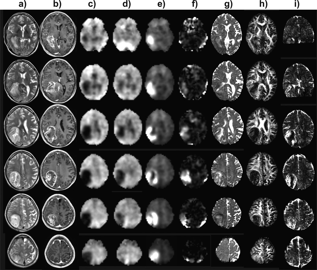
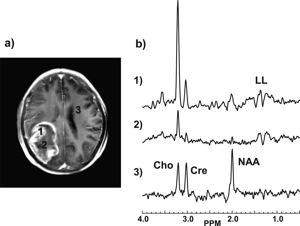
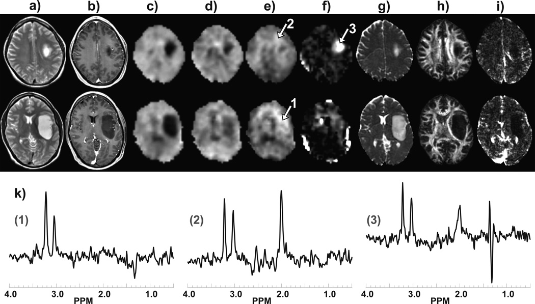
Introduction: Accurate grading of cerebral glioma using conventional structural imaging techniques remains challenging due to the relatively poor sensitivity and specificity of these methods. The purpose of this study was to evaluate the relative sensitivity and specificity of structural magnetic resonance imaging and MR measurements of perfusion, diffusion, and whole-brain spectroscopic parameters for glioma grading.
Methods: Fifty-six patients with radiologically suspected untreated glioma were studied with T1- and T2-weighted MR imaging, dynamic contrast-enhanced MR imaging, diffusion tensor imaging, and volumetric whole-brain MR spectroscopic imaging. Receiver-operating characteristic analysis was performed using the relative cerebral blood volume (rCBV), apparent diffusion coefficient, fractional anisotropy, and multiple spectroscopic parameters to determine optimum thresholds for tumor grading and to obtain the sensitivity, specificity, and positive and negative predictive values for identifying high-grade gliomas. Logistic regression was performed to analyze all the parameters together.
Results: The rCBV individually classified glioma as low and high grade with a sensitivity and specificity of 100 and 88 %, respectively, based on a threshold value of 3.34. On combining all parameters under consideration, the classification was achieved with 2 % error and sensitivity and specificity of 100 and 96 %, respectively.
Conclusion: Individually, CBV measurement provides the greatest diagnostic performance for predicting glioma grade; however, the most accurate classification can be achieved by combining all of the imaging parameters.
Figures
References
-
- Chiang IC, Kuo YT, Lu CY, et al. Distinction between high-grade gliomas and solitary metastases using peritumoral 3-T magnetic resonance spectroscopy, diffusion, and perfusion imagings. Neuroradiology. 2004;46:619–627. - PubMed
-
- Catalaa I, Henry R, Dillon WP, et al. Perfusion, diffusion and spectroscopy values in newly diagnosed cerebral gliomas. NMR Biomed. 2006;19:463–475. - PubMed
-
- Fink JR, Carr RB, Matsusue E, et al. Comparison of 3 Tesla proton MR spectroscopy, MR perfusion and MR diffusion for distinguishing glioma recurrence from posttreatment effects. J Magn Reson Imaging. 2012;35:56–63. - PubMed
-
- Lee EJ, Brugge K, Mikulis D, et al. Diagnostic value of peritumoral minimum apparent diffusion coefficient for differentiation of glioblastoma multiforme from solitary metastatic lesions. AJR. 2011;196:71–76. - PubMed
Publication types
MeSH terms
Grants and funding
LinkOut - more resources
Full Text Sources
Other Literature Sources
Medical
