. 2013 Mar;20(3):317-25.
doi: 10.1038/nsmb.2499. Epub 2013 Feb 3.

Acetylation limits 53BP1 association with damaged chromatin to promote homologous recombination

Affiliations

Acetylation limits 53BP1 association with damaged chromatin to promote homologous recombination

Jiangbo Tang et al. Nat Struct Mol Biol. 2013 Mar.

Abstract

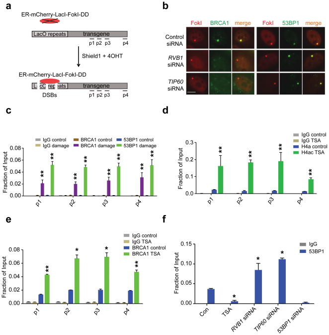
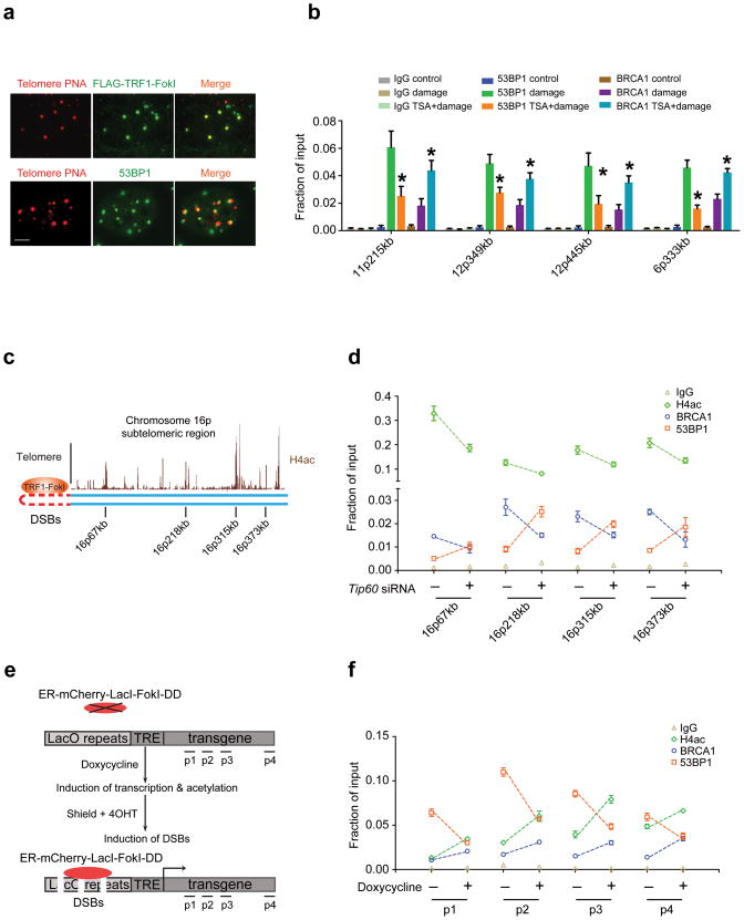
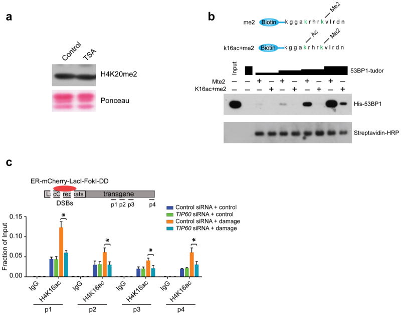
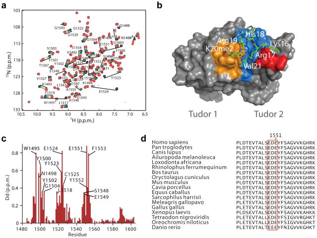
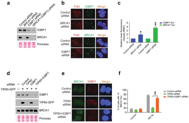
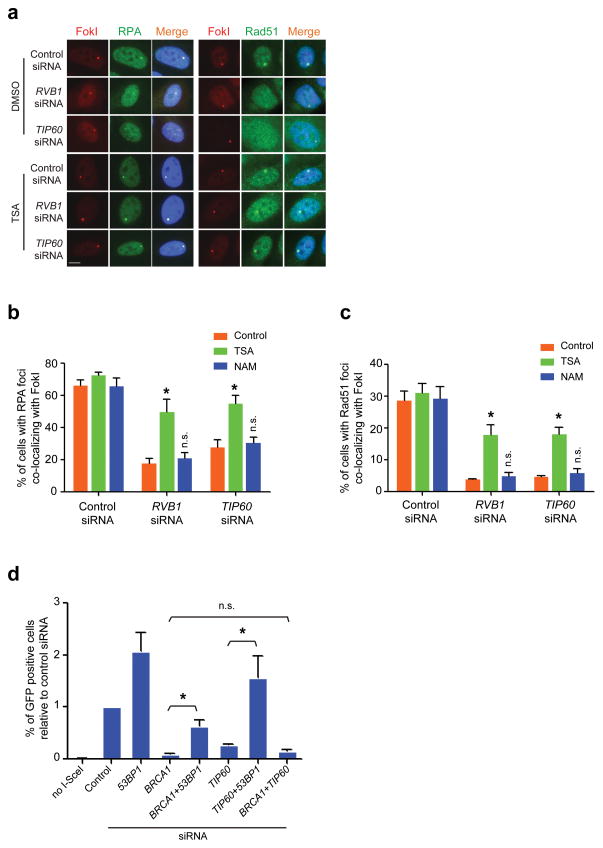
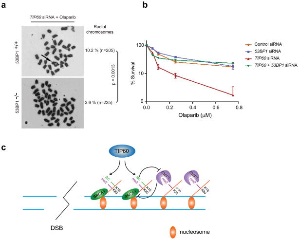
The pathogenic sequelae of BRCA1 mutation in human and mouse cells are mitigated by concomitant deletion of 53BP1, which binds histone H4 dimethylated at Lys20 (H4K20me2) to promote nonhomologous end joining, suggesting that a balance between BRCA1 and 53BP1 regulates DNA double strand-break (DSB) repair mechanism choice. Here we document that acetylation is a key determinant of this balance. TIP60 acetyltransferase deficiency reduced BRCA1 at DSB chromatin with commensurate increases in 53BP1, whereas HDAC inhibition yielded the opposite effect. TIP60-dependent H4 acetylation diminished 53BP1 binding to H4K20me2 in part through disruption of a salt bridge between H4K16 and Glu1551 in the 53BP1 Tudor domain. Moreover, TIP60 deficiency impaired homologous recombination and conferred sensitivity to PARP inhibition in a 53BP1-dependent manner. These findings demonstrate that acetylation in cis to H4K20me2 regulates relative BRCA1 and 53BP1 DSB chromatin occupancy to direct DNA repair mechanism.


Conflict of interest statement

COMPETING FINANCIAL INTERESTS

The authors declare no competing financial interests.

Figures

Figure 1
Figure 1. TIP60 and HDAC inhibition differentially impact competition between BRCA1 and 53BP1 for localization to DSBs
(a) Schematic of inducible DSB generation by mCherry-LacI-FokI at an integrated reporter transgene in U2OS cells (U2OS-DSB-reporter). ChIP-qPCR primer sets are shown as p1–p4. (b) Immunofluorescence (IF) was performed at 5 hours after induction of DSBs by mCherry-LacI-FokI in the U2OS-DSB-reporter cells transfected with the indicated siRNA. Scale bar, 10 μm. (c) ChIP-qPCR was performed with an antibody to 53BP1 or BRCA1 in the U2OS-DSB-reporter cell line with (Damage) or without (Control) induction of DSBs by mCherry-LacI-FokI. Error bars represent S.E.M. ** p<0.005. (d) ChIP-qPCR was performed with antibody to H4ac in the U2OS-DSB-reporter cell line after treatment with DMSO (Control) or 0.5 μM TSA. Induction of DSBs by mCherry-LacI-FokI was included in all conditions tested. Error bars represent S.E.M. ** p<0.005. (e) ChIP-qPCR was performed with antibody to BRCA1. Cells were treated with DMSO (Control) or 0.5 μM TSA for 5 hours and DSBs were induced by mCherry-LacI-FokI. Error bars represent S.E.M. * indicates p<0.05, ** p<0.005. (f) ChIP-qPCR was performed with antibody to 53BP1 in the U2OS-DSB-reporter cells (locus p4). DSBs were induced at 48–72 hours after siRNA transfection or 5 hours after TSA (0.5 μM) administration. Error bars represent S.E.M. * indicates p<0.05.
Figure 2
Figure 2. DNA damage and transcription dependent acetylation regulates BRCA1 and 53BP1 DSB occupancy
(a) TRF1-FokI colocalizes with telomeres and 53BP1 foci are observed adjacent to telomeric signals as shown by immuno-FISH using peptide nucleic acid probes against telomeres (Telomere PNA). Scale bar, 10 μm. (b) ChIP using antibodies to BRCA1 and 53BP1 at different subtelomeric regions in 293T cells with (Damage) or without (Control) expression of TRF1-FokI. Treatment with TSA is indicated. The chromosome location and distance from the telomere is indicated at each qPCR primer set. Error bars represent standard error of the mean (S.E.M). * indicates p<0.05. (c) Schematic of the subtelomeric region of 16p denoting histone H4ac pattern (UCSC genome browser) and positions of ChIP-qPCR primer sets. (d) ChIP was performed at regions of high H4ac as indicated in c in 293T cells expressing TRF1-FokI following transfection of control or TIP60 siRNA. (e) Schematic of DSB induction in transcriptionally inactive or active chromatin. Transcription is induced with doxycycline prior to DSB induction. (f) ChIP was performed in the presence or absence of doxycycline as described in e. Induction of DSBs by mCherry-LacI-FokI was included in all conditions tested following the addition of doxycycline. Error bars represent S.E.M. * indicates p<0.05.
Figure 3
Figure 3. Histone H4K16 acetylation impairs 53BP1 Tudor domain interaction with H4K20me2
(a) Immunoblot (IB) was performed on acid-eluted histones from U2OS cells that had been treated for 5 hours with 0.5 μM TSA. (b) Biotinylated H4 peptide encompassing amino acids 12–25 with the relevant methylated and acetylated lysine residues highlighted in green (top panel) were used to pull down increasing amounts of His-53BP1-Tudor. IB was performed as indicated (bottom panel). (c) ChIP was performed using antibodies to H4K16ac in the absence (Control) or presence (Damage) of mCherry-LacI-FokI-induced DSBs in cells transfected with either control or TIP60 specific siRNA.
Figure 4
Figure 4. Solution structure of 53BP1-Tudor in association with a methylated H4 peptide
(a) Changes in the 1H-15N HSQC correlation spectrum of 53BP1-Tudor upon addition of increasing amounts of H4K20me2 peptide. The 53BP1 correlation signals affected by the interaction with the peptide are labeled. (b) NMR structure of 53BP1-Tudor in complex with a histone H4K20me2 peptide. The peptide (aa 14-27) is in stick represention with only key interacting residues shown. 53BP1-Tudor is in gray surface representation with its methyllysine binding cage highlighted in orange. Important hydrophobic residues for which intermolecular NOEs were detected are shown in blue. The red area corresponds to 53BP1 Asp1550 and 2 Glu1551. (c) Histogram of 1H-15N chemical shift changes Δδ=(δH)2+(δN/5)2 for the backbone amide atoms and Trp1495 HN and Asn1498 HN of 53BP1-Tudor observed upon saturation with the H4K20me2 peptide. The signals of Ser1496 and Gly1499 disappeared completely after titration with H4K20me2. Backbone HN signals of Trp1495, Ser1497 and Asn1498 were not detected, even in the absence of peptide. (d) Amino acid sequence alignment of 53BP1 from various vertebrate species illustrating conservation of the 1549–1551 acidic patch in the second Tudor domain.
Figure 5
Figure 5. 53BP1 knockdown restores BRCA1 localization to DSBs
(a) IB with cell lysates prepared from cells treated with siRNAs as indicated. (b) Representative images of 53BP1 (upper panel) or BRCA1 (lower panel) at mCherry-LacI-FokI-induced DSBs in cells transfected with the indicated siRNA. Scale bar, 10 μm. (c), Quantification of relative mean fluorescence intensity (RMFI) from b. RMFI was calculated from 200 cells and 3 independent experiments. Error bars represent S.E.M. * indicates p<0.05, ** p<0.005. (d), IB with cell lysates prepared from TIP60-GFP expressing cells treated with siRNAs as indicated. (e) Representative images of BRCA1 and 53BP1 co-staining following fixation at 5 hours after 2 Gy IR treatment of U2OS cells that were transfected with the indicated siRNA. Scale bar, 10 μm. (f) Quantification of percentage of cells possessing greater than 5 BRCA1 IRIF per cell from e. Error bars represent S.E.M. ** indicates p<0.005.
Figure 6
Figure 6. HDACi or 53BP1 deficiency restores end resection and homology directed DSB repair in TIP60 deficient cells
(a) IF was performed with antibodies to RPA or Rad51 in siRNA transfected U2OS-DSB-reporter cells that were incubated with either DMSO control or TSA (0.5 μM). Scale bar, 10 μm. (b, c) Quantification of RPA and Rad51 DSB localization from a. Analysis was performed on 200 cells per group in at least 2 independent experiments. Error bars represent S.E.M. * p < 0.05, n.s. not significant. (d) Homology directed repair of I-SceI DSBs in siRNA transfected U2OS DR-GFP reporter cells as assessed by FACS for percent GFP positive cells. *p<0.05, n.s. = not significant.
Figure 7
Figure 7. 53BP1 promotes genomic instability and sensitivity to PARPi in the absence of TIP60
(a) Representative metaphase images from 53BP1+/+ and 53BP1−/− MEFs following knockdown of TIP60 and Olaparib treatment. Significance was calculated using Fisher’s exact test. (b) Percentage survival following Olaparib administration in U2OS cells that had been transfected with the indicated siRNAs. (c) Model depicting that TIP60 -dependent acetylation limits 53BP1 binding to H4K20Me2 when present on the same histone H4 tail. TIP60 deficiency would result in hypoacetylated H4K16 and a 53BP1-dependent block of BRCA1 DSB chromatin association.

Comment in

  • DNA damage: limiting 53BP1.
    Du Toit A. Du Toit A. Nat Rev Mol Cell Biol. 2013 Mar;14(3):132. doi: 10.1038/nrm3532. Epub 2013 Feb 13. Nat Rev Mol Cell Biol. 2013. PMID: 23403718 No abstract available.

References

    1. Bunting SF, et al. 53BP1 inhibits homologous recombination in Brca1-deficient cells by blocking resection of DNA breaks. Cell. 2010;141:243–54. - PMC - PubMed
    1. Bouwman P, et al. 53BP1 loss rescues BRCA1 deficiency and is associated with triple-negative and BRCA-mutated breast cancers. Nat Struct Mol Biol. 2010;17:688–95. - PMC - PubMed
    1. Chapman JR, Taylor MR, Boulton SJ. Playing the end game: DNA double-strand break repair pathway choice. Mol Cell. 2012;47:497–510. - PubMed
    1. Xu X, et al. Genetic interactions between tumor suppressors Brca1 and p53 in apoptosis, cell cycle and tumorigenesis. Nat Genet. 2001;28:266–71. - PubMed
    1. Botuyan MV, et al. Structural basis for the methylation state-specific recognition of histone H4-K20 by 53BP1 and Crb2 in DNA repair. Cell. 2006;127:1361–73. - PMC - PubMed

Publication types

MeSH terms

Associated data