. 2013 May;31(5):992-1000.
doi: 10.1002/stem.1338.

Lgr5-EGFP marks taste bud stem/progenitor cells in posterior tongue

Affiliations

Lgr5-EGFP marks taste bud stem/progenitor cells in posterior tongue

Karen K Yee et al. Stem Cells. 2013 May.

Abstract

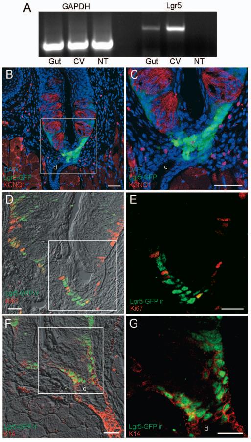
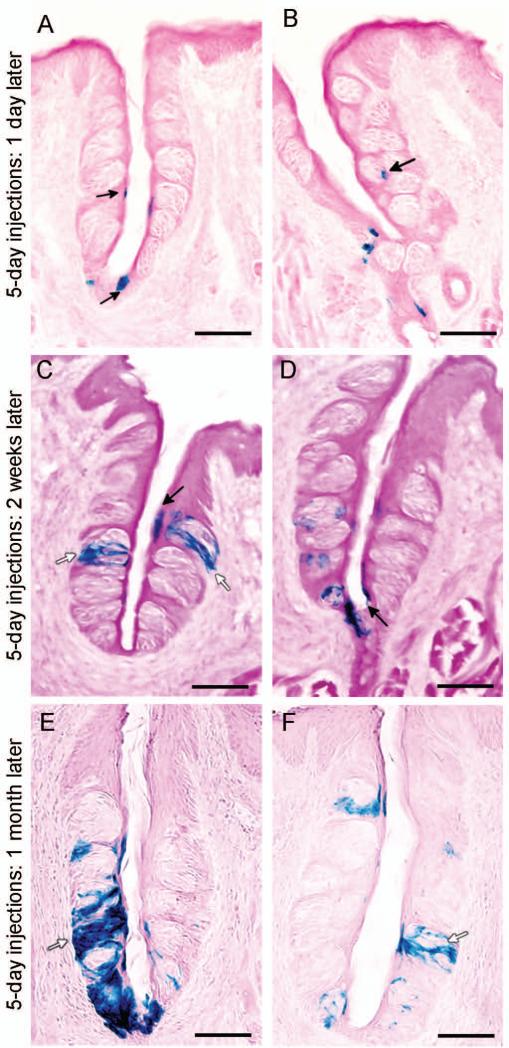
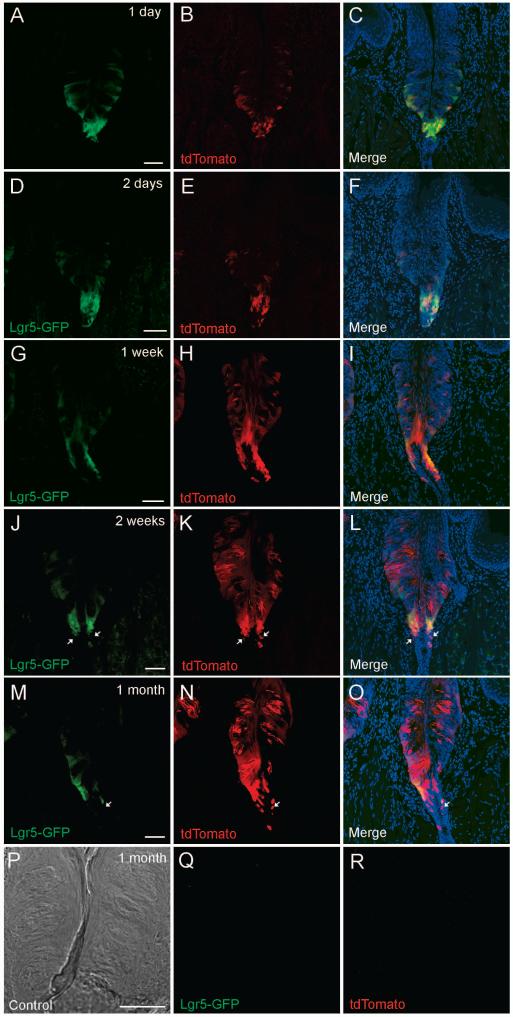
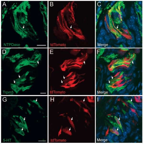
Until recently, reliable markers for adult stem cells have been lacking for many regenerative mammalian tissues. Lgr5 (leucine-rich repeat-containing G-protein-coupled receptor 5) has been identified as a marker for adult stem cells in intestine, stomach, and hair follicle; Lgr5-expressing cells give rise to all types of cells in these tissues. Taste epithelium also regenerates constantly, yet the identity of adult taste stem cells remains elusive. In this study, we found that Lgr5 is strongly expressed in cells at the bottom of trench areas at the base of circumvallate (CV) and foliate taste papillae and weakly expressed in the basal area of taste buds and that Lgr5-expressing cells in posterior tongue are a subset of K14-positive epithelial cells. Lineage-tracing experiments using an inducible Cre knockin allele in combination with Rosa26-LacZ and Rosa26-tdTomato reporter strains showed that Lgr5-expressing cells gave rise to taste cells, perigemmal cells, along with self-renewing cells at the bottom of trench areas at the base of CV and foliate papillae. Moreover, using subtype-specific taste markers, we found that Lgr5-expressing cell progeny include all three major types of adult taste cells. Our results indicate that Lgr5 may mark adult taste stem or progenitor cells in the posterior portion of the tongue.


Figures

Fig. 1
Fig. 1. Lgr5 is expressed in circumvallate papilla
A) RT-PCR demonstrates Lgr5 expression in gut (positive control) and circumvallate (CV) papilla taste tissue but not in the surrounding non-taste (NT) epithelium. The GAPDH RT-PCR controls confirm equivalent template amounts from gut, CV, and NT cDNA. B-G) Confocal images of Lgr5+ cells in CV papilla. B and C) The Lgr5-GFP transgene was detected by intrinsic fluorescence (green). The strongest GFP signal is at the bottom of the trench area below the CV papilla and adjacent to the opening of the ducts of Von Ebner's glands. A weaker GFP signal is found in cells at the base of the taste buds immediately below and between the mature taste cells (red), which were immunodetected with a KCNQ1 antibody. D and E) Dual immunostaining shows that only some Lgr5+ cells (green, anti-GFP antibody) at the bottom of the CV trench area are also immunoreactive for proliferation marker Ki67 (red, anti-Ki67). F and G) Dual immunostaining shows that all Lgr5+ cells (green, anti-GFP) at the bottom of the CV trench area are also immunoreactive for K14 (red, anti-K14). All images are compressed confocal z-stacks (~5μm thickness). d, duct of Von Ebner's gland. Scale bars: B=80 μm, C=10 μm, and D-G=20μm.
Fig. 2
Fig. 2. Lgr5-expressing cells give rise to mature taste cells
Tamoxifen-induced Lgr5-Cre generates β-galactosidase activity to mark cells from the Lgr5+ lineage. A and B) Representative images of β-galactosidase-stained taste cells (blue) of the Lgr5+ lineage present in the trench at the base of the circumvallate papilla, at the taste pore, and at the basal layer of the taste buds (arrows), one day after 5-days of tamoxifen induction of Lgr5-Cre. C and D) β-Galactosidase-stained taste cells (blue) of the Lgr5+ lineage mark both intragemmal (within the taste bud; white arrows) and perigemmal (surrounding the taste bud; black arrows) cells, two weeks after 5-days of tamoxifen induction of Lgr5-Cre. E and F) β-Galactosidase-stained taste cells (blue) of the Lgr5+ lineage mark many intragemmal taste cells (white arrows), one month after 5-days of tamoxifen induction of Lgr5-Cre. Scale bars: A-F=40 μm.
Fig. 3
Fig. 3. Lgr5-expressing cells give rise to mature taste cells and cells in the associated ducts
Tamoxifen-induced Lgr5-Cre generates tdTomato fluorescent protein (red) to mark cells from the Lgr5+ lineage. Representative confocal images of the circumvallate papilla, one day (A-C), two days (D-F), one week (G-I), two weeks (J-L) and one month (M-O) after a single tamoxifen induction of Lgr5-Cre. Images in the left column (A, D, G, J, M) show Lgr5-GFP intrinsic fluorescence (green) at the indicated times. Images in the middle column (B, E, H, K, N) show tdTomato+ (red) cells. The right column (C, F, I, L, O) shows the merged images. Note that increasing numbers of tdTomato+ intragemmal taste cells and perigemmal cells are generated over time after a single tamoxifen induction. At all dates examined after a single tamoxifen induction many Lgr5-GFP+ cells are also tdTomato+ in both the trench area and at the base of the taste buds. A few cells (white arrows) that are tdTomato+ but not Lgr5-GFP+ are present in the ductal area under the trench two weeks or 1 month after a single tamoxifen induction. P-R) A transmitted light image of a circumvallate section (P) of an Lgr5+/+, Rosa26-tdTomato+/- mouse (negative control littermate 1 month after a single tamoxifen induction) shows no EGFP (Q) or tdTomato (R) fluorescence when scanned at the same confocal settings. All images are compressed confocal z-stacks (~8μm thickness). Scale bar: 20 μm.
Fig. 4
Fig. 4. Lgr5+ stem/progenitor cells generate all three types of taste bud cells
Mature taste bud cells marked by Lgr5-Cre-generated tdTomato 1 month after tamoxifen induction were stained with markers for each of the three types of taste cells. A-C) tdTomato (red) is co-expressed in type I taste cells with NTPDase2 (pseudo-colored green). D-F) Lgr5-Cre-generated tdTomato (red) is co-expressed in type II taste receptor cells with Trpm5 (pseudo-colored green). G-I) Lgr5-Cre-generated tdTomato (red) is co-expressed in type III taste receptor cells with serotonin (pseudo-colored green). Arrows show colocalization of tdTomato and taste specific markers. All images are compressed confocal z-stacks (~3 μm thickness). Scale bars: 10 μm.

Similar articles

Cited by

References

    1. Bachmanov AA, Beauchamp GK. Taste receptor genes. Annu Rev Nutr. 2007;27:389–414. - PMC - PubMed
    1. Beidler LM, Smallman RL. Renewal of cells within taste buds. J Cell Biol. 1965;27:263–272. - PMC - PubMed
    1. Cohn ZJ, Kim A, Huang L, et al. Lipopolysaccharide-induced inflammation attenuates taste progenitor cell proliferation and shortens the life span of taste bud cells. BMC Neurosci. 2010;11:72. - PMC - PubMed
    1. Hamamichi R, Asano-Miyoshi M, Emori Y. Taste bud contains both short-lived and long-lived cell populations. Neuroscience. 2006;141:2129–2138. - PubMed
    1. Okubo T, Clark C, Hogan BL. Cell lineage mapping of taste bud cells and keratinocytes in the mouse tongue and soft palate. Stem Cells. 2009;27:442–450. - PMC - PubMed

Publication types

MeSH terms

Substances