Review
. 2012 Dec 15;125(Pt 24):5917-26.
doi: 10.1242/jcs.093732. Epub 2013 Feb 1.

At the leading edge of three-dimensional cell migration

Affiliations
Review

At the leading edge of three-dimensional cell migration

Ryan J Petrie et al. J Cell Sci. .

Abstract

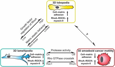
Cells migrating on flat two-dimensional (2D) surfaces use actin polymerization to extend the leading edge of the plasma membrane during lamellipodia-based migration. This mode of migration is not universal; it represents only one of several mechanisms of cell motility in three-dimensional (3D) environments. The distinct modes of 3D migration are strongly dependent on the physical properties of the extracellular matrix, and they can be distinguished by the structure of the leading edge and the degree of matrix adhesion. How are these distinct modes of cell motility in 3D environments related to each other and regulated? Recent studies show that the same type of cell migrating in 3D extracellular matrix can switch between different leading edge structures. This mode-switching behavior, or plasticity, by a single cell suggests that the apparent diversity of motility mechanisms is integrated by a common intracellular signaling pathway that governs the mode of cell migration. In this Commentary, we propose that the mode of 3D cell migration is governed by a signaling axis involving cell-matrix adhesions, RhoA signaling and actomyosin contractility, and that this might represent a universal mechanism that controls 3D cell migration.


Figures

Fig. 1.
Fig. 1.
Mechanosensing of matrix rigidity by RhoA, ROCK and myosin II and its potential regulation by Rho–Rac crosstalk. The RhoA–ROCK–myosin-II signaling axis is capable of sensing changes in the structure of the extracellular matrix and responding to it by increasing actomyosin contractility. The actin-binding motor protein myosin II maintains a low level of tension on actin fibers that are coupled to the extracellular matrix through cell–matrix adhesions. This basal tension enables myosin II to respond to changes in matrix rigidity or elastic behavior by increasing the tension on cell–matrix adhesions to activate the GEFs GEF-H1 and LARG. These GEFs activate RhoA, which in turn activates ROCK to increase the phosphorylation of myosin light chain (MLC), thereby further increasing myosin II activity and actomyosin contractility. This mechanical feedback loop can increase integrin clustering and adhesion maturation, and might increase intracellular pressure and plasma membrane tension to prevent lamellipodia formation and promote lobopodia- and bleb-based motility. Crosstalk between Rac1 and RhoA signaling potentially regulates the mechanosensing of matrix rigidity and the mode of 3D cell migration. During mesenchymal (lamellipodial) melanoma cell migration, NEDD9 forms a complex with the Rac1 GEF DOCK3 to activate Rac1 and suppress MLC phosphorylation through the Rac effector WAVE2, thereby suppressing amoeboid migration. Conversely, during amoeboid migration RhoA-dependent ROCK signaling can activate the Rac1 GAPs ARHGAP22 and FilGAP to inactivate Rac1 and suppresses mesenchymal migration in melanoma and carcinoma cells, respectively.
Fig. 2.
Fig. 2.
Pseudopodium identity can define the mode of 2D and 3D cell migration. There is a wide diversity in the types of pseudopodia, or protrusions, that are used to extend the leading edge during cell migration on 2D surfaces and in 3D extracellular matrix. The type of pseudopodium can be used to define a specific mode of cell motility. (A) Lamellipodia-based migration is used by cells that migrate on 2D glass (upper and middle panels) and in a 3D collagen matrix (lower panel). Lamellipodia are thin fan-shaped protrusions enriched in F-actin and actin-binding proteins such as cortactin. (B) Lobopodia are blunt cylindrical protrusions that might be driven by intracellular pressure rather than actin polymerization. They are classically associated with giant amoeba (lower panel), but are also formed by metazoan cells migrating in linear elastic 3D material, such as cell-derived matrix (CDM) (upper panel). (C) Round amoeboid migration of cancer cells in 3D collagen comprises at least three distinct modes of migration that are characterized by multiple small blebs (upper panel), large hemispherical blebs (middle panel), or an actin-enriched leading edge (lower panel). (D) Rhizopodia and axopodia are pseudopodia used by certain protozoa to migrate and feed. LM, lamellipodium; LB, lobopodium. Broken white arrows indicate the direction of migration.
Fig. 3.
Fig. 3.
Mechanical control of the mode of 3D cell migration. Three modes of 3D metazoan cell motility can be uniquely identified based on only two characteristics: their degree of cell–matrix adhesion and their requirement for RhoA, ROCK and myosin II activity. 3D lobopodia-based motility is associated with robust cell–matrix adhesion and requires RhoA signaling. Reducing RhoA activity, through soluble signaling factors or changes in the elastic behavior of the 3D matrix, causes adherent cells to undergo a transition to 3D lamellipodia-based motility. Cancer cells switch to the rounded low adhesion, high contractility mode of amoeboid cancer cell motility upon inhibition of protease activity or upon modulation of Rho GTPase crosstalk. Cells that use lamellipodia might have low membrane tension; therefore increased actomyosin contractility could elevate membrane tension and prevent lamellipodia formation during both lobopodial and round amoeboid cancer cell migration. It remains to be determined how cells capable of lobopodia-based migration transition to round bleb-based motility (indicated by the question mark).

References

    1. Abercrombie M., Heaysman J. E., Pegrum S. M. (1970a). The locomotion of fibroblasts in culture. I. Movements of the leading edge. Exp. Cell Res. 59, 393–398 10.1016/0014-4827(70)90646-4 - DOI - PubMed
    1. Abercrombie M., Heaysman J. E., Pegrum S. M. (1970b). The locomotion of fibroblasts in culture. II. “Ruffling”. Exp. Cell Res. 60, 437–444 10.1016/0014-4827(70)90537-9 - DOI - PubMed
    1. Ahn J., Sanz–Moreno V., Marshall C. J. (2012). The metastasis gene NEDD9 product acts through integrin β3 and Src to promote mesenchymal motility and inhibit amoeboid motility. J. Cell Sci. 125, 1814–1826 10.1242/jcs.101444 - DOI - PubMed
    1. Albrecht–Buehler G. (1980). Autonomous movements of cytoplasmic fragments. Proc. Natl. Acad. Sci. USA 77, 6639–6643 10.1073/pnas.77.11.6639 - DOI - PMC - PubMed
    1. Allen R. D., Allen N. S. (1978). Cytoplasmic streaming in amoeboid movement. Annu. Rev. Biophys. Bioeng. 7, 469–495 10.1146/annurev.bb.07.060178.002345 - DOI - PubMed

Publication types